


Nori Bovine TGFb1 ELISA Kit

Price range: $508.00 through $916.00
This ELISA kit is for quantification of TGFb1 in bovine. This is a quick ELISA assay that reduces time to 50% compared to the conventional method, and the entire assay only takes 3 hours. This assay employs the quantitative sandwich enzyme immunoassay technique and uses biotin-streptavidin chemistry to improve the performance of the assays. An antibody specific for TGFb1 has been pre-coated onto a microplate. Standards and samples are pipetted into the wells and any TGFb1 present is bound by the immobilized antibody. After washing away any unbound substances, a detection antibody specific for TGFb1 is added to the wells. Following wash to remove any unbound antibody reagent, a detection reagent is added. After intensive wash a substrate solution is added to the wells and color develops in proportion to the amount of TGFb1 bound in the initial step. The color development is stopped, and the intensity of the color is measured.
Alternative names for TGFb1: Transforming growth factor beta 1
This product is for laboratory research use only not for diagnostic and therapeutic purposes or any other purposes.
- Description
- How Elisa Works
- Product Citations (19)
- Reviews (0)
Description
Nori Bovine TGFb1 ELISA Kit Summary
Alternative names for TGFb1: Transforming growth factor beta 1
Alternative names for bovine: cattle, cow, bull
| Assay Type | Solid Phase Sandwich ELISA |
| Format | 96-well Microplate or 96-Well Strip Microplate |
| Method of Detection | Colorimetric |
| Number of Targets Detected | 1 |
| Target Antigen Accession Number | P18341 |
| Assay Length | 3 hours |
| Quantitative/Semiquantitative | Quantitative |
| Sample Type | Plasma, Serum, Cell Culture, Urine, Cell/Tissue Lysates, Synovial Fluid, BAL, |
| Recommended Sample Dilution (Plasma/Serum) | No dilution for sample <ULOQ; sufficient dilution for samples >ULOQ |
| Sensitivity | 6 pg/mL |
| Detection Range | 31-2000 pg/mL |
| Specificity | Bovine TGFb1 |
| Cross-Reactivity | < 0.5% cross-reactivity observed with available related molecules, < 50% cross-species reactivity observed with species tested. |
| Interference | No significant interference observed with available related molecules |
| Storage/Stability | 4 ºC for up to 6 months |
| Usage | For Laboratory Research Use Only. Not for diagnostic or therapeutic use. |
| Additional Notes | The kit allows for use in multiple experiments. |
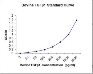
Standard Curve
Kit Components
1. Pre-coated 96-well Microplate
2. Biotinylated Detection Antibody
3. Streptavidin-HRP Conjugate
4. Lyophilized Standards
5. TMB One-Step Substrate
6. Stop Solution
7. 20 x PBS
8. Assay Buffer
Other Materials Required but not Provided:
1. Microplate Reader capable of measuring absorption at 450 nm
2. Log-log graph paper or computer and software for ELISA data analysis
3. Precision pipettes (1-1000 µl)
4. Multi-channel pipettes (300 µl)
5. Distilled or deionized water
Protocol Outline
1. Prepare all reagents, samples and standards as instructed in the datasheet.
2. Add 100 µl of Standard or samples to each well and incubate 1 h at RT.
3. Add 100 µl of Working Detection Antibody to each well and incubate 1 h at RT.
4. Add 100 µl of Working Streptavidin-HRP to each well and incubate 20 min at RT.
5. Add 100 µl of Substrate to each well and incubate 5-30 min at RT.
6. Add 50 µl of Stop Solution to each well and read at 450 nm immediately.
Background:
TGFβ1 (transforming growth factor beta 1) was first identified in human platelets as a protein with a molecular mass of 25 kilodaltons with a potential role in wound healing (1). TGFB1, TGFB2, and TGFB3 all function through the same receptor signaling systems. They are members of the large TGFβ superfamily. TGFβ proteins are highly pleiotropic cytokines that regulate processes such as immune function, proliferation and epithelial mesenchymal transition (2-4). It was later characterized as a large protein precursor (containing 390 amino acids) that was proteolytically processed to produce a mature peptide of 112 amino acids (5). TGFβ activation from latency is controlled both spatially and temporally, by multiple pathways that include actions of proteases such as plasmin and MMP9, and/or by thrombospondin 1 or selected integrins (5, 6). Although different isoforms of TGFβ are naturally associated with their own distinct LAPs, the TGFβ1 LAP is capable of complexing with, and inactivating, all other Equine TGFβ isoforms and those of most other species (7). Mutations within the LAP are associated with Camurati Engelmann disease, a rare sclerosing bone dysplasia characterized by inappropriate presence of active TGFβ1 (8).
References
- Assoian R, et al. (1983). J Biol Chem 258 (11): 7155.
- Dunker, N. and K. Krieglstein (2000) Eur. J. Biochem.267:6982.
- Wahl, S.M. (2006) Immunol. Rev. 213:213.
- Chang, H. et al. (2002) Endocr. Rev. 23:787.
- Derynck, R. et al. (1985) Nature 316:701.
- Oklu, R. and R. Hesketh (2000) Biochem. J. 352:601.
- Miller, D.M. et al. (1992) Mol. Endocrinol. 6:694.
- Janssens, K. et al. (2003) J. Biol. Chem. 278:7718.

Product Citations
Nori® TGFb1 ELISA Kit Citations
Since 2014, updated on 5-Apr-26
- Khalil Y. (2013) Profiling immune responses in diary calves experimentally infected with Mycobacterium avium Subsp. Paratuberculosis (Map). MS Thesis, University of Calgary (Canada). Product used and cited: Nori bovine TGFβ1 ELISA kit.
- Zucca E et al. (2016) Evaluation of amniotic mesenchymal cell derivatives on cytokine production in equine alveolar macrophages: an in vitro approach to lung inflammation. Stem Cell Research and Therapy 7:137. Impact factor: 4.731. Product used and cited: Nori Equine TNF-α, IL-6 and TGFβ1 ELISA kits.
- Sun Y et al. (2017) Oral administration of saccharomyces boulardii alters duodenal morphology, enzymatic activity and cytokine production response in broiler chickens. Animal Sci J, 88:1204-1211. Impact factor: 1.325. Products used and cited: Nori Chicken IL-6, TNFa, IL-10 and TGFb ELISA kits.
- Lee Ju and Kim GH (2017) Calcium-deficient hydroxyapatite/collagen/platelet-rich plasma scaffold with controlled release function for hard tissue regeneration. ACS Biomater Sci Eng. DOI: 10.1021/ascbiomaterials.7b00640. Impact factor: 3.234. Products used and cited: Nori Guinea Pig TGF-b1 and PDGF ELISA Kits.
- Jones AK et al. (2017) Gestational restricted- and over-feeding promote maternal and offspring inflammatory response that are distinct and dependent on diet on sheep. Biology of Reproduction doi:10.1093/biolre/iox174. Impact factor: 3.714. Products used and cited: Nori Sheep BMP2/HGF/TGFb1 ELISA Kits.
- Min Y-J et al. (2018) Effects of eco-friendly multi-enzyme on diarrhea and immune response of weaned pigs. Korean J Org Agric 26(1):151-161. Products used and cited: Nori Porcine TNF-alpha, TGF-beta 1, and CRP ELISA Kits.
- Leander, AAK (2017) A comparative analysis of equine mesenchymal cells and dermal fibroblasts. The University of Waikato, Master of Philosophy Thesis. Products used and cited: Nori Equine TNF-alpha, IL-1 beta, IL-6, TGF-beta 1, IL-10 and PGE2 ELISA Kits.
- Tsai H-C et al. (2019) A mini-pig model for evaluating the efficiency of autologous platelet patches on induced acute full thickness wound healing. BMC Vet Res 15:191. Impact factor: 1.80. Produce used and cited: Nori Porcine TGFb1 ELISA Kit.
- USDA Grant proposal (2018) Recovery of biologically active compounds from byproducts of Louisiana aquaculture for dietary supplement and biomedical applications. Louisiana State University. http://portal.nifa.usda.gov/crtsprojectpages/1017853. Products cited: Nori Equine TGFβ1, IGF-1, PDGF, EGF ELISA Kits.
- Yang Q (2019) Equine Hoof Stratum internum K14+CD105+progenitor cells: culture, characterization, and model of epithelial to mesenchymal transition. PhD Thesis, Louisiana State Univ. Products used and cited: Nori Equine TGFβ1 ELISA Kit.
- Marteles D et al. (2019) Effects of allergen-specific immunotherapy on peripheral blood regulatory T cells and serum concentrations of cytokines and immunoglobulins in horses with allergic dermatitis. Intl Immunopharmacol 74:105674. Impact factor: 1.846. Products used and cited: Nori Equine TGFβ1, IL-10, and IgE ELISA Kits.
- Gonzalez I et al. (2019) Difference in blood parameters associated to stress response between Chilean rodeo horses and Chilean urban working horses. J Equine Vet Sci 73:110-4. Impact factor: 0.35. Products used and cited: Nori Equine TGFβ1 ELISA Kit.
- Lee, JJ et al. (2020) Effects of dietary protease on immune response of weaned pigs. J Anim Sci Technol 62(2): 174-9. Impact factor: NA. Products used and cited: Nori Porcine TNFa, TGFb1 and CRP ELISA Kits (GR106162, GR106126, GR113093).
- Canciello A et al. (2020) Progesterone prolongs viability and anti-inflammatory functions of explanted preterm ovine amniotic membrane. Frontiers in Bioengineering and Biotechnology 8 Article 135. frontiersin.org. Impact factor: 3.52. Products used and cited: Nori Sheep IL-4, IL-10 and TGFb1 ELISA Kits (GR106451, GR106454, GR106127).
- Kang et al. (2021) Effects of dietary inactivated probiotics on growth performance and immune responses of weaned pigs. JASTorg/10.5187/jast.2021.e44. Impact factor: 1.685. Products used and cited: Nori Porcine TNFα, TGFβ1 and CRP ELISA Kits.
- Rostami F et al. (2021) Effects of scrophularia striata hydroalcoholic extract in comparison to salinomycin on growth performance, intestinal health and immunity in broiler chickens following a mixed-species Eimeria challenge. Vet Parasitol 293:109417. org/10.1016/j.vetpar.2021.109417. Impact factor: 2.009. Products used and cited: Nori Chicken IgM, IgG, TGFβ1 and IL-6 ELISA Kits.
- Haspeslagh M et al. (2021) Limited added value of negative pressure wound intention healing in a non-contaminated and contaminated equine distal limb wound model. Equine Vet J org/10.1111/evj.13487. Impact factor: 2.888. Products used and cited: Nori Equine TGF beta 1 ELISA Kits
- Song M et al. (2022) Modification of Gut Microbiota and Immune Responses via Dietary Protease in Soybean Meal-Based Protein Diets. J Microbiol Biotechnol 32 (7):885-891. Impact factor: 3.1. Products used and cited: Nori Porcine CRP, TNFa and TGFb1 ELISA Kits
- Karabinowska-Malocha A et al. (2023) Link between fibrosis-specific biomarkers and interstitial fibrosis in hypertrophic cardiomyopathy. Kardiologia Polska Impact factor: 3.3 https://doi.org/10.33963/kp.a2023.0103. Products used and cited: Nori Human Gal-3, TGFb1 ELISA Kit.
Be the first to review “Nori Bovine TGFb1 ELISA Kit”
You must be logged in to post a review.
Reviews
There are no reviews yet.