


Nori Bovine EGF ELISA Kit

Price range: $508.00 through $916.00
This ELISA kit is for quantification of EGF in bovine. This is a quick ELISA assay that reduces time to 50% compared to the conventional method, and the entire assay only takes 3 hours. This assay employs the quantitative sandwich enzyme immunoassay technique and uses biotin-streptavidin chemistry to improve the performance of the assays. An antibody specific for EGF has been pre-coated onto a microplate. Standards and samples are pipetted into the wells and any EGF present is bound by the immobilized antibody. After washing away any unbound substances, a detection antibody specific for EGF is added to the wells. Following wash to remove any unbound antibody reagent, a detection reagent is added. After intensive wash a substrate solution is added to the wells and color develops in proportion to the amount of EGF bound in the initial step. The color development is stopped, and the intensity of the color is measured.
Alternative names for EGF: Epidermal growth factor
This product is for laboratory research use only not for diagnostic and therapeutic purposes or any other purposes.
- Description
- How Elisa Works
- Product Citations (2)
- Reviews (0)
Description
Nori Bovine EGF ELISA Kit Summary
Alternative names for EGF: Epidermal growth factor
Alternative name for bovine: cow, cattle, bull
| Assay Type | Solid Phase Sandwich ELISA |
| Format | 96-well Microplate or 96-Well Strip Microplate |
| Method of Detection | Colorimetric |
| Number of Targets Detected | 1 |
| Target Antigen Accession Number | XP_024849546.1 |
| Assay Length | 3 hours |
| Quantitative/Semiquantitative | Quantitative |
| Sample Type | Plasma, Serum, Cell Culture, Urine, Cell/Tissue Lysates, Synovial Fluid, BAL, |
| Recommended Sample Dilution (Plasma/Serum) | No dilution for sample <ULOQ; sufficient dilution for samples >ULOQ |
| Sensitivity | 1.2 pg/mL |
| Detection Range | 6.3-400 pg/mL |
| Specificity | Bovine EGF |
| Cross-Reactivity | < 0.5% cross-reactivity observed with available related molecules, < 50% cross-species reactivity observed with species tested. |
| Interference | No significant interference observed with available related molecules |
| Storage/Stability | 4 ºC for up to 6 months |
| Usage | For Laboratory Research Use Only. Not for diagnostic or therapeutic use. |
| Additional Notes | The kit allows for use in multiple experiments. |
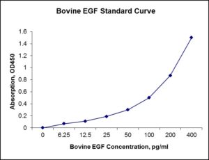
Standard Curve
Kit Components
1. Pre-coated 96-well Microplate
2. Biotinylated Detection Antibody
3. Streptavidin-HRP Conjugate
4. Lyophilized Standards
5. TMB One-Step Substrate
6. Stop Solution
7. 20 x PBS
8. Assay Buffer
Other Materials Required but not Provided:
1. Microplate Reader capable of measuring absorption at 450 nm
2. Log-log graph paper or computer and software for ELISA data analysis
3. Precision pipettes (1-1000 µl)
4. Multi-channel pipettes (300 µl)
5. Distilled or deionized water
Protocol Outline
1. Prepare all reagents, samples and standards as instructed in the datasheet.
2. Add 100 µl of Standard or samples to each well and incubate 1 h at RT.
3. Add 100 µl of Working Detection Antibody to each well and incubate 1 h at RT.
4. Add 100 µl of Working Streptavidin-HRP to each well and incubate 20 min at RT.
5. Add 100 µl of Substrate to each well and incubate 5-30 min at RT.
6. Add 50 µl of Stop Solution to each well and read at 450 nm immediately.
Background:
Epidermal growth factor (EGF) is a growth factor that stimulates cell growth, proliferation, and differentiation by binding to its receptor EGFR. Human EGF is a 6045-Da protein[1] with 53 amino acid residues and three intramolecular disulfide bonds.[2] EGF results in cellular proliferation, differentiation, and survival.[3] EGF is a low-molecular-weight polypeptide first purified from the mouse submandibular gland, but since then found in many human tissues including submandibular gland, parotid gland. Salivary EGF, which seems also regulated by dietary inorganic iodine, also plays an important physiological role in the maintenance of oro-esophageal and gastric tissue integrity. The biological effects of salivary EGF include healing of oral and gastroesophageal ulcers, inhibition of gastric acid secretion, stimulation of DNA synthesis as well as mucosal protection from intraluminal injurious factors such as gastric acid, bile acids, pepsin, and trypsin and to physical, chemical and bacterial agents.[4] EGF acts by binding with high affinity to epidermal growth factor receptor (EGFR) on the cell surface. This stimulates ligand-induced dimerization,[5]activating the intrinsic protein-tyrosine kinase activity of the receptor (see the second diagram). The tyrosine kinase activity, in turn, initiates a signal transduction cascade that results in a variety of biochemical changes within the cell – a rise in intracellular calcium levels, increased glycolysis and protein synthesis, and increases in the expression of certain genes including the gene for EGFR – that ultimately lead to DNA synthesis and cell proliferation.[6]
References
- Harris RC, et al. (2003). Experimental Cell Research 284 (1): 2–13.
- Carpenter G, et al. (1990). The Journal of Biological Chemistry 265 (14): 7709–12.
- Herbst RS (2004). Intl J Radiation Oncology, Biology, Physics 59 (2 Suppl): 21–6.
- Venturi S, et al. (2009). Nutrition and Health 20 (2): 119–134.
- Dawson JP, et al. (2005). Mol. Cell. Biol. 25 (17): 7734–42.
- Fallon JH, et al. (1984). Science 224 (4653): 1107–9.

Product Citations
- USDA Grant proposal (2018) Recovery of biologically active compounds from byproducts of Louisiana aquaculture for dietary supplement and biomedical applications. Louisiana State University. http://portal.nifa.usda.gov/crtsprojectpages/1017853. Products cited: Nori Equine TGFβ1, IGF-1, PDGF, EGF ELISA Kits.
- Jeong W et al. (2022) Therapeutic Effects of Amnion-Conjugated Chitosan-Alginate Membranes on Diabetic Wounds in an Induced Diabetic Swine Model: An In Vitro and In Vivo Study. Archives of Plastic Surgery 49(2):258-265. Impact factor: 1.5. Products used and cited: Nori Bovine EGF, FGF-2, KGF ELISA Kit. Article
Be the first to review “Nori Bovine EGF ELISA Kit”
You must be logged in to post a review.
Reviews
There are no reviews yet.