


Nori Canine PDGF-D ELISA Kit

Price range: $508.00 through $916.00
This ELISA kit is for quantification of PDGF-DD in dog. This is a quick ELISA assay that reduces time to 50% compared to the conventional method, and the entire assay only takes 3 hours. This assay employs the quantitative sandwich enzyme immunoassay technique and uses biotin-streptavidin chemistry to improve the performance of the assays. An antibody specific for PDGF-DD has been pre-coated onto a microplate. Standards and samples are pipetted into the wells and any PDGF-DD present is bound by the immobilized antibody. After washing away any unbound substances, a detection antibody specific for PDGF-DD is added to the wells. Following wash to remove any unbound antibody reagent, a detection reagent is added. After intensive wash a substrate solution is added to the wells and color develops in proportion to the amount of PDGF-DD bound in the initial step. The color development is stopped, and the intensity of the color is measured.
Alternative names for PDGF-DD: Platelet-derived growth factor D, PDGFD
This product is for Laboratory Research Use Only not for diagnostic and therapeutic purposes or any other purposes.
- Description
- How Elisa Works
- Product Citation (0)
- Reviews (0)
Description
Nori Canine PDGF-DD ELISA Kit Summary
Alternative names for PDGF-DD: Platelet-derived growth factor D, PDGFD
Alternative name for canine: dog
| Assay Type | Solid Phase Sandwich ELISA |
| Format | 96-well Microplate or 96-Well Strip Microplate |
| Method of Detection | Colorimetric |
| Number of Targets Detected | 1 |
| Target Antigen Accession Number | A0A8C0MSL5 |
| Assay Length | 3 hours |
| Quantitative/Semiquantitative | Quantitative |
| Sample Type | Plasma, Serum, Cell Culture, Urine, Cell/Tissue Lysates, Synovial Fluid, BAL, |
| Recommended Sample Dilution (Plasma/Serum) | No dilution for sample <ULOQ; sufficient dilution for samples >ULOQ |
| Sensitivity | 5 pg/mL |
| Detection Range | 25-1600 pg/mL |
| Specificity | Canine PDGF-DD |
| Cross-Reactivity | < 0.5% cross-reactivity observed with available related molecules, < 50% cross-species reactivity observed with species tested. |
| Interference | No significant interference observed with available related molecules |
| Storage/Stability | 4 ºC for up to 6 months |
| Usage | For Laboratory Research Use Only. Not for diagnostic or therapeutic use. |
| Additional Notes | The kit allows for use in multiple experiments. |
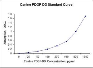
Standard Curve
Kit Components
1. Pre-coated 96-well Microplate
2. Biotinylated Detection Antibody
3. Streptavidin-HRP Conjugate
4. Lyophilized Standards
5. TMB One-Step Substrate
6. Stop Solution
7. 20 x PBS
8. Assay Buffer
Other Materials Required but not Provided:
1. Microplate Reader capable of measuring absorption at 450 nm
2. Log-log graph paper or computer and software for ELISA data analysis
3. Precision pipettes (1-1000 µl)
4. Multi-channel pipettes (300 µl)
5. Distilled or deionized water
Protocol Outline
1. Prepare all reagents, samples and standards as instructed in the datasheet.
2. Add 100 µl of Standard or samples to each well and incubate 1 h at RT.
3. Add 100 µl of Working Detection Antibody to each well and incubate 1 h at RT.
4. Add 100 µl of Working Streptavidin-HRP to each well and incubate 20 min at RT.
5. Add 100 µl of Substrate to each well and incubate 5-30 min at RT.
6. Add 50 µl of Stop Solution to each well and read at 450 nm immediately.
Background:
Platelet-derived growth factor D is a protein that is encoded by the PDGFD gene and is a member of the platelet-derived growth factor family.[1] The four members of this family are mitogenic factors for cells of mesenchymal origin and are characterized by a core motif of eight cysteines, seven of which are found in PDGF-D. This gene product only forms homodimers and, therefore, does not dimerize with the other three family members. It differs from alpha and beta members of this family in having an unusual N-terminal domain, the CUB domain. PDGF-D is a specific protease-activated ligand for the PDGF beta-receptor.[2] PDGF-D is expressed in many tissues such as kidney and eye.[3,4] PDGF-D is activated by urokinase plasminogen activator in prostate carcinoma cells.[5] Inducible PDGF-D expression by angiotensin II and hydrogen peroxide involves transcriptional regulation by Ets-1 and Sp1.[6] PDGF-D and its PDGF receptor beta (PDGFR-beta) are expressed in colorectal cancer.[7] PDGF-D overexpression is an independent predictor of platinum-based chemotherapy resistance and it may also be a potential biomarker for targeted therapy and poor prognosis.[8]
References
- LaRochelle WJ, et al. (2001). Nat. Cell Biol. 3 (5): 517–21.
- Bergsten E, et al. (2001). Nat. Cell Biol. 3 (5): 512–6.
- Changsirikulchai S, et al. (2003). Kidney Int. 62 (6): 2043–54.
- Ray S, et al. (2005). J. Biol. Chem. 280(9): 8494–502.
- Ustach CV, Kim HR (2005) Mol. Cell. Biol. 25 (14): 6279–88.
- Liu MY, et al. (2006) Blood. 107 (6): 2322–9.
- Olsen RS, et al. (2019) Cancer Invest. 37 (2), 99-112.
- Zhang,M., et al. (2018) Int. J. Gynecol. Cancer 28 (2), 323-331.

Be the first to review “Nori Canine PDGF-D ELISA Kit”
You must be logged in to post a review.
Reviews
There are no reviews yet.