



















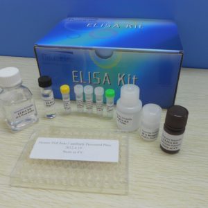
Nori Zebrafish HSP27 ELISA Kit
Price range: $508.00 through $916.00
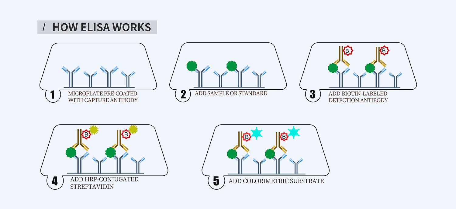
This ELISA kit is for quantification of HSP27 in zebrafish. This is a quick ELISA assay that reduces time to 50% compared to the conventional method, and the entire assay only takes 3 hours. This assay employs the quantitative sandwich enzyme immunoassay technique and uses biotin-streptavidin chemistry to improve the performance of the assays. An antibody specific for HSP27 has been pre-coated onto a microplate. Standards and samples are pipetted into the wells and any HSP27 present is bound by the immobilized antibody. After washing away any unbound substances, a detection antibody specific for HSP27 is added to the wells. Following wash to remove any unbound antibody reagent, a detection reagent is added. After intensive wash a substrate solution is added to the wells and color develops in proportion to the amount of HSP27 bound in the initial step. The color development is stopped, and the intensity of the color is measured.
Alternative names for HSP27: Heat shock protein 27, HSP beta-1, HSPB1
This product is for Laboratory Research Use Only not for diagnostic and therapeutic purposes or any other purposes.
- Description
- How Elisa Works
- Documents
- Product Citation (0)
- Reviews (0)
Description
Nori Zebrafish HSP27 ELISA Kit Summary
Alternative names for HSP27: Heat shock protein 27, HSP beta-1, HSPB1
Alternative names for zebrafish: Danio rerio
| Assay Type | Solid Phase Sandwich ELISA |
| Format | 96-well Microplate or 96-Well Strip Microplate |
| Method of Detection | Colorimetric |
| Number of Targets Detected | 1 |
| Target Antigen Accession Number | A0ACM8QCU4 |
| Assay Length | 3 hours |
| Quantitative/Semiquantitative | Quantitative |
| Sample Type | Plasma, Serum, Cell Culture, Urine, Cell/Tissue Lysates, Synovial Fluid, BAL, |
| Recommended Sample Dilution (Plasma/Serum) | No dilution for sample <ULOQ; sufficient dilution for samples >ULOQ |
| Sensitivity | 6 pg/mL |
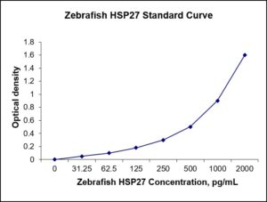
| Detection Range | 31.25-2000 pg/mL |
| Specificity | Zebrafish HSP27 |
| Cross-Reactivity | < 0.5% cross-reactivity observed with available related molecules, < 50% cross-species reactivity observed with species tested. |
| Interference | No significant interference observed with available related molecules |
| Storage/Stability | 4 ºC for up to 6 months |
| Usage | For Laboratory Research Use Only. Not for diagnostic or therapeutic use. |
| Additional Notes | The kit allows for use in multiple experiments. |
Standard Curve
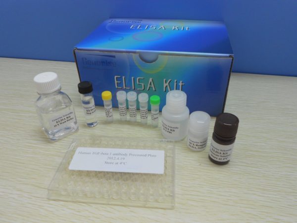
Kit Components
1. Pre-coated 96-well Microplate
2. Biotinylated Detection Antibody
3. Streptavidin-HRP Conjugate
4. Lyophilized Standards
5. TMB One-Step Substrate
6. Stop Solution
7. 20 x PBS
8. Assay Buffer
Other Materials Required but not Provided:
1. Microplate Reader capable of measuring absorption at 450 nm
2. Log-log graph paper or computer and software for ELISA data analysis
3. Precision pipettes (1-1000 µl)
4. Multi-channel pipettes (300 µl)
5. Distilled or deionized water
Protocol Outline
1. Prepare all reagents, samples and standards as instructed in the datasheet.
2. Add 100 µl of Standard or samples to each well and incubate 1 h at RT.
3. Add 100 µl of Working Detection Antibody to each well and incubate 1 h at RT.
4. Add 100 µl of Working Streptavidin-HRP to each well and incubate 20 min at RT.
5. Add 100 µl of Substrate to each well and incubate 5-30 min at RT.
6. Add 50 µl of Stop Solution to each well and read at 450 nm immediately.
Background:
Heat shock protein 27 (Hsp27) also known as HSP beta-1 (HSPB1) is a protein that is encoded by the HSPB1gene.[1] Hsp27 is a chaperone of the sHsp (small HSP) group among ubiquitin, α-crystallin, Hsp20 and others. The common functions of sHsps are chaperone activity, thermotolerance, inhibition of apoptosis, regulation of cell development, and cell differentiation. They also take part in signal transduction. Hsp27 appears in many cell types, especially all types of muscle cells. It is located mainly in the cytosol, but also in the perinuclear region, endoplasmatic reticulum, and nucleus. It is overexpressed during different stages of cell differentiation and development. This suggests an essential role for Hsp27 in the differentiation of tissues. The main function of Hsp27 is to provide thermotolerance in vivo, cytoprotection, and support of cell survival under stress conditions. Hsp27 interacts with the outer mitochondrial membranes and interferes with the activation of cytochrome c/Apaf-1/dATP complex and therefore inhibits the activation of procaspase-9.[2] The phosphorylated form of Hsp27 inhibits Daxx apoptotic protein and prevents the association of Daxx with Fas and Ask1.[3] Hsp27 interacts with actin and intermediate filaments. It prevents the formation of non-covalent filament/filament interactions of the intermediate filaments and protects actin filaments from fragmentation. It also preserves the focal contacts fixed at the cell membrane.[2] Another function of Hsp27 is the activation of the proteasome. It speeds up the degradation of irreversibly denatured proteins and junk proteins by binding to ubiquitinated proteins and to the 26S proteasome. Hsp27 enhances the activation of the NF-κB pathway, that controls a lot of processes, such as cell growth and inflammatory and stress responses.[4] The cytoprotective properties of Hsp27 result from its ability to modulate reactive oxygen species and to raise glutathione levels.
References
- Carper SW, et al. (November 1990). Nucleic Acids Res. 18 (21): 6457.
- Sarto C, et al. (April 2000). Electrophoresis 21 (6): 1218–26.
- Charette SJ, et al. (October 2000). Mol. Cell. Biol. 20 (20): 7602–12.
- Parcellier A, Schmitt E, Gurbuxani S, Seigneurin-Berny D, Pance A, Chantôme A, Plenchette S, Khochbin S, Solary E, Garrido C (August 2003). Mol. Cell. Biol. 23 (16): 5790–802.
Be the first to review “Nori Zebrafish HSP27 ELISA Kit”
You must be logged in to post a review.
Reviews
There are no reviews yet.