


















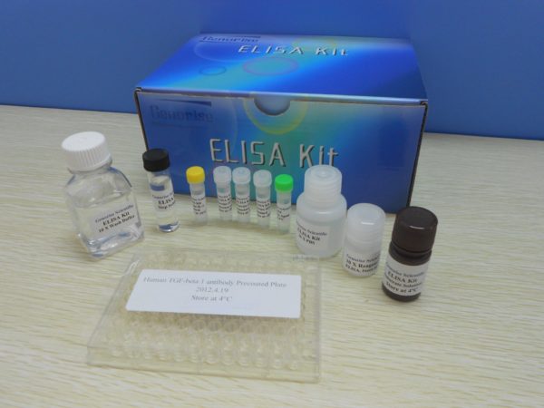
Nori Human AMH ELISA Kit
Price range: $508.00 through $916.00
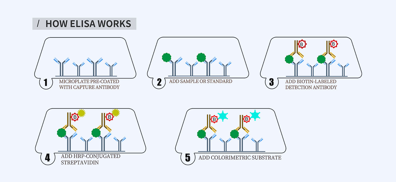
This ELISA kit is for quantification of AMH in human. This is a quick ELISA assay that reduces time to 50% compared to the conventional method, and the entire assay only takes 3 hours. This assay employs the quantitative sandwich enzyme immunoassay technique and uses biotin-streptavidin chemistry to improve the performance of the assays. An antibody specific for AMH has been pre-coated onto a microplate. Standards and samples are pipetted into the wells and any AMH present is bound by the immobilized antibody. After washing away any unbound substances, a detection antibody specific for AMH is added to the wells. Following wash to remove any unbound antibody reagent, a detection reagent is added. After intensive wash a substrate solution is added to the wells and color develops in proportion to the amount of AMH bound in the initial step. The color development is stopped, and the intensity of the color is measured.
Alternative names for AMH: Anti-Müllerian hormone, Müllerian-inhibiting hormone, MIH, MIF
This product is for laboratory research use only not for diagnostic and therapeutic purposes or any other purposes.
- Description
- How Elisa Works
- Documents
- Product Citations
- Reviews (0)
Description
Nori Human AMH ELISA Kit Summary
Alternative names for AMH: Anti-Müllerian hormone, Müllerian-inhibiting hormone, MIH, MIF
| Assay Type | Solid Phase Sandwich ELISA |
| Format | 96-well Microplate or 96-Well Strip Microplate |
| Method of Detection | Colorimetric |
| Number of Targets Detected | 1 |
| Target Antigen Accession Number | P03971 |
| Assay Length | 3 hours |
| Quantitative/Semiquantitative | Quantitative |
| Sample Type | Plasma, Serum, Cell Culture, Urine, Cell/Tissue Lysates, Synovial Fluid, BAL, |
| Recommended Sample Dilution (Plasma/Serum) | No dilution for sample <ULOQ; sufficient dilution for samples >ULOQ |
| Sensitivity | 25 pg/mL |
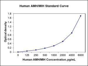
| Detection Range | 125-6000 pg/mL |
| Specificity | Human AMH |
| Cross-Reactivity | < 0.5% cross-reactivity observed with available related molecules, < 50% cross-species reactivity observed with species tested. |
| Interference | No significant interference observed with available related molecules |
| Storage/Stability | 4 ºC for up to 6 months |
| Usage | For Laboratory Research Use Only. Not for diagnostic or therapeutic use. |
| Additional Notes | The kit allows for use in multiple experiments. |
Standard Curve
Kit Components
1. Pre-coated 96-well Microplate
2. Biotinylated Detection Antibody
3. Streptavidin-HRP Conjugate
4. Lyophilized Standards
5. TMB One-Step Substrate
6. Stop Solution
7. 20 x PBS
8. Assay Buffer
Other Materials Required but not Provided:
1. Microplate Reader capable of measuring absorption at 450 nm
2. Log-log graph paper or computer and software for ELISA data analysis
3. Precision pipettes (1-1000 µl)
4. Multi-channel pipettes (300 µl)
5. Distilled or deionized water
Protocol Outline
1. Prepare all reagents, samples and standards as instructed in the datasheet.
2. Add 100 µl of Standard or samples to each well and incubate 1 h at RT.
3. Add 100 µl of Working Detection Antibody to each well and incubate 1 h at RT.
4. Add 100 µl of Working Streptavidin-HRP to each well and incubate 20 min at RT.
5. Add 100 µl of Substrate to each well and incubate 5-30 min at RT.
6. Add 50 µl of Stop Solution to each well and read at 450 nm immediately.
Background:
Anti-Müllerian hormone (AMH), also known as Müllerian-inhibiting hormone (MIH), is a glycoprotein hormone structurally related to inhibin and activin from the transforming growth factor beta superfamily, whose key roles are in growth differentiation and folliculogenesis. AMH is activated by SOX9 in the Sertoli cells of the male fetus.[1] Its expression inhibits the development of the female reproductive tract, or Müllerian ducts, in the male embryo, thereby arresting the development of fallopian tubes, uterus, and upper vagina.[2] AMH expression is critical to sex differentiation at a specific time during fetal development, and appears to be tightly regulated by nuclear receptor SF-1, transcription GATA factors, sex-reversal gene DAX1, and follicle-stimulating hormone (FSH).[3][4] Mutations in both the AMH gene and the type II AMH receptor have been shown to cause the persistence of Müllerian derivatives in males that are otherwise normally masculinized. AMH is also a product of granulosa cells of the preantral and small antral follicles in women. As such, AMH is only present in the ovary until menopause. Production of AMH regulates folliculogenesis by inhibiting recruitment of follicles from the resting pool in order to select for the dominant follicle, after which the production of AMH diminishes.[5] AMH can also serve as a biomarker for relative size of the ovarian reserve.[6] In bovine, AMH can be used for selection of females in multi-ovulatory embryo transfer programs by predicting the number of antral follicles developed to ovulation.[7] AMH can also be used as a marker for ovarian dysfunction, such as in women with polycystic ovary syndrome (PCOS).
References
- Taguchi O, et al. (1984). Developmental Biology. 106 (2): 394–8.
- Panidis D, et al. (2011). Medical Hypotheses. 77 (4): 649–53.
- Shen WH, et al. (1994) Cell. 77 (5): 651–61.
- Viger RS, et al. (1998). Development. 125 (14): 2665–75.
- Kollmann Z, et al. (2015). Reproductive Biology and Endocrinology. 13: 21.
- Weenen C, et al. (2004). Molecular Human Reproduction. 10 (2): 77–83.
- Rico C, et al. (2011). Biology of Reproduction. 84 (3): 560–71.
Product Citations
Be the first to review “Nori Human AMH ELISA Kit”
You must be logged in to post a review.
Reviews
There are no reviews yet.