


Nori Equine Insulin ELISA Kit

Price range: $461.00 through $832.00
This ELISA kit is for quantification of insulin in equine. This is a quick ELISA assay that reduces time to 50% compared to the conventional method, and the entire assay only takes 3 hours. This assay employs the quantitative sandwich enzyme immunoassay technique and uses biotin-streptavidin chemistry to improve the performance of the assays. An antibody specific for INS has been pre-coated onto a microplate. Standards and samples are pipetted into the wells and any INS present is bound by the immobilized antibody. After washing away any unbound substances, a detection antibody specific for INS is added to the wells. Following wash to remove any unbound antibody reagent, a detection reagent is added. After intensive wash a substrate solution is added to the wells and color develops in proportion to the amount of INS bound in the initial step. The color development is stopped, and the intensity of the color is measured.
Alternative names for insulin: INS
This product is for Laboratory Research Use Only not for diagnostic and therapeutic purposes or any other purposes.
- Description
- How Elisa Works
- Product Citation (0)
- Reviews (0)
Description
Nori Equine Insulin ELISA Kit Summary
Alternative names for insulin: INS
Alternative names for equine: horse
| Assay Type | Solid Phase Sandwich ELISA |
| Format | 96-well Microplate or 96-Well Strip Microplate |
| Method of Detection | Colorimetric |
| Number of Targets Detected | 1 |
| Target Antiben Accession Number | na |
| Assay Length | 3 hours |
| Quantitative/Semiquantitative | Quantitative |
| Sample Type | Plasma, Serum, Cell Culture, Urine, Cell/Tissue Lysates, Synovial Fluid, BAL, |
| Recommended Sample Dilution (Plasma/Serum) | No dilution for sample <ULOQ; sufficient dilution for samples >ULOQ |
| Sensitivity | 0.6 uIU/mL |
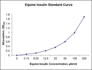
| Detection Range | 3.13-200 uIU/mL |
| Specificity | Equine insulin |
| Cross-Reactivity | < 0.5% cross-reactivity observed with available related molecules, < 50% cross-species reactivity observed with species tested. |
| Interference | No significant interference observed with available related molecules |
| Storage/Stability | 4 ºC for up to 6 months |
| Usage | For Laboratory Research Use Only. Not for diagnostic or therapeutic use. |
| Additional Notes | The kit allows for use in multiple experiments. |
Standard Curve
Kit Components
1. Pre-coated 96-well Microplate
2. Biotinylated Detection Antibody
3. Streptavidin-HRP Conjugate
4. Lyophilized Standards
5. TMB One-Step Substrate
6. Stop Solution
7. 20 x PBS
8. Assay Buffer
Other Materials Required but not Provided:
1. Microplate Reader capable of measuring absorption at 450 nm
2. Log-log graph paper or computer and software for ELISA data analysis
3. Precision pipettes (1-1000 µl)
4. Multi-channel pipettes (300 µl)
5. Distilled or deionized water
Protocol Outline
1. Prepare all reagents, samples and standards as instructed in the datasheet.
2. Add 100 µl of Standard or samples to each well and incubate 1 h at RT.
3. Add 100 µl of Working Detection Antibody to each well and incubate 1 h at RT.
4. Add 100 µl of Working Streptavidin-HRP to each well and incubate 20 min at RT.
5. Add 100 µl of Substrate to each well and incubate 5-30 min at RT.
6. Add 50 µl of Stop Solution to each well and read at 450 nm immediately.
Background:
Insulin is a peptide hormone produced by beta cells of the pancreatic islets and is the main anabolic hormone of the body. It regulates the metabolism of carbohydrates, fats and protein by promoting the absorption of glucose from the blood into liver, fat and skeletal muscle cells. In these tissues the absorbed glucose is converted into either glycogen via glycogenesis or fats (triglycerides) via lipogenesis, or, in the case of the liver, into both. Glucose production and secretion by the liver is strongly inhibited by high concentrations of insulin in the blood.[1] Circulating insulin also affects the synthesis of proteins in a wide variety of tissues. It promotes the conversion of small molecules in the blood into large molecules inside the cells. Low insulin levels in the blood have the opposite effect by promoting widespread catabolism, especially of reserve body fat. Beta cells are sensitive to blood sugar levels so that they secrete insulin into the blood in response to high level of glucose; and inhibit secretion of insulin when glucose levels are low.[2] Insulin enhances glucose uptake and metabolism in the cells, thereby reducing blood sugar level. The secretion of insulin and glucagon into the blood in response to the blood glucose concentration is the primary mechanism of glucose homeostasis.[2] Decreased or absent insulin activity results in diabetes mellitus, a condition of high blood sugar level (hyperglycaemia). There are two types of the disease. In type 1 diabetes mellitus, the beta cells are destroyed by an autoimmune reaction so that insulin can no longer be synthesized or be secreted into the blood. In type 2 diabetes mellitus, the destruction of beta cells is less pronounced than in type 1 diabetes, and is not due to an autoimmune process. Instead, there is an accumulation of amyloid in the pancreatic islets, which likely disrupts their anatomy and physiology.[2] The pathogenesis of type 2 diabetes is not well understood but reduced population of islet beta-cells, reduced secretory function of islet beta-cells that survive, and peripheral tissue insulin resistance are known to be involved. Type 2 diabetes is characterized by increased glucagon secretion which is unaffected by, and unresponsive to the concentration of blood glucose. But insulin is still secreted into the blood in response to the blood glucose.[2] As a result, glucose accumulates in the blood.
References
- Sonksen P, Sonksen J (2000). British Journal of Anaesthesia. 85(1): 69–79.
- Koeslag JH, et al. (2003). The Journal of Physiology. 549 (Pt 2): 333–46.

Be the first to review “Nori Equine Insulin ELISA Kit”
You must be logged in to post a review.
Reviews
There are no reviews yet.