


Nori Goat IgE ELISA Kit

Price range: $508.00 through $916.00
This ELISA kit is for quantification of IgE in goat. This is a quick ELISA assay that reduces time to 50% compared to the conventional method, and the entire assay only takes 3 hours. This assay employs the quantitative sandwich enzyme immunoassay technique and uses biotin-streptavidin chemistry to improve the performance of the assays. An antibody specific for IgE has been pre-coated onto a microplate. Standards and samples are pipetted into the wells and any IgE present is bound by the immobilized antibody. After washing away any unbound substances, a detection antibody specific for IgE is added to the wells. Following wash to remove any unbound antibody reagent, a detection reagent is added. After intensive wash a substrate solution is added to the wells and color develops in proportion to the amount of IgE bound in the initial step. The color development is stopped, and the intensity of the color is measured.
Alternative names for IgE: Immunoglobulin E
This product is for laboratory research use only not for diagnostic and therapeutic purposes or any other purposes.
- Description
- How Elisa Works
- Product Citations (3)
- Reviews (0)
Description
Nori Goat IgE ELISA Kit Summary
Alternative names for IgE: Immunoglobulin E
Alternative names for goat: ovine, sheep, lamb, caprine
| Assay Type | Solid Phase Sandwich ELISA |
| Format | 96-well Microplate or 96-Well Strip Microplate |
| Method of Detection | Colorimetric |
| Number of Targets Detected | 1 |
| Target Antigen Accession Number | na |
| Assay Length | 3 hours |
| Quantitative/Semiquantitative | Quantitative |
| Sample Type | Plasma, Serum, Cell Culture, Urine, Cell/Tissue Lysates, Synovial Fluid, BAL, |
| Recommended Sample Dilution (Plasma/Serum) | No dilution for sample <ULOQ; sufficient dilution for samples >ULOQ |
| Sensitivity | 120 pg/mL |
| Detection Range | 0.625-40 ng/mL |
| Specificity | Goat IgE |
| Cross-Reactivity | < 0.5% cross-reactivity observed with available related molecules, < 50% cross-species reactivity observed with species tested. |
| Interference | No significant interference observed with available related molecules |
| Storage/Stability | 4 ºC for up to 6 months |
| Usage | For Laboratory Research Use Only. Not for diagnostic or therapeutic use. |
| Additional Notes | The kit allows for use in multiple experiments. |
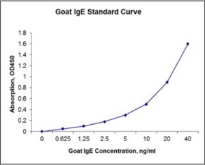
Standard Curve
Kit Components
1. Pre-coated 96-well Microplate
2. Biotinylated Detection Antibody
3. Streptavidin-HRP Conjugate
4. Lyophilized Standards
5. TMB One-Step Substrate
6. Stop Solution
7. 20 x PBS
8. Assay Buffer
Other Materials Required but not Provided:
1. Microplate Reader capable of measuring absorption at 450 nm
2. Log-log graph paper or computer and software for ELISA data analysis
3. Precision pipettes (1-1000 µl)
4. Multi-channel pipettes (300 µl)
5. Distilled or deionized water
Protocol Outline
1. Prepare all reagents, samples and standards as instructed in the datasheet.
2. Add 100 µl of Standard or samples to each well and incubate 1 h at RT.
3. Add 100 µl of Working Detection Antibody to each well and incubate 1 h at RT.
4. Add 100 µl of Working Streptavidin-HRP to each well and incubate 20 min at RT.
5. Add 100 µl of Substrate to each well and incubate 5-30 min at RT.
6. Add 50 µl of Stop Solution to each well and read at 450 nm immediately.
Background:
Immunoglobulin E (IgE) is a kind of antibody (or immunoglobulin (Ig) “isotype“) that has only been found in mammals. Monomers of IgE consist of two heavy chains (ε chain) and two light chains, with the ε chain containing 4 Ig-like constant domains (Cε1-Cε4). IgE’s main function is immunity to parasites such as helminths[1] like Schistosoma mansoni, Trichinella spiralis, and Fasciola hepatica.[2] IgE is utilized during immune defense against certain protozoan parasites such as Plasmodium falciparum. IgE also has an essential role in type I hypersensitivity,[3] which manifests various allergic diseases, such as allergic asthma, most types of sinusitis, allergic rhinitis, food allergies, and specific types of chronic urticaria and atopic dermatitis. IgE also plays a pivotal role in responses to allergens, such as: anaphylactic drugs, bee stings, and antigen preparations used in desensitization immunotherapy. Although IgE is typically the least abundant isotype—blood serum IgE levels in a normal (“non-atopic“) individual are only 0.05% of the Ig concentration,[4] compared to 75% for the IgGs at 10 mg/ml, which are the isotypes responsible for most of the classical adaptive immune response—it is capable of triggering the most powerful inflammatory reactions. IgE primes the IgE-mediated allergic response by binding to Fc receptors found on the surface of mast cells and basophils. IgE may play an important role in the immune system’s recognition of cancer,[5] in which the stimulation of a strong cytotoxic response against cells displaying only small amounts of early cancer markers would be beneficial. IgE is known to be elevated in various autoimmune disorders such as Lupus(SLE), Rheumatoid Arthritis(RA) & psoriasis, and is theorized to be of pathogenetic importance in RA and SLE by eliciting a hypersensitivity reaction.[6]
References
- Erb KJ (2007). Eur. J. Immunol. 37 (5): 1170–3.
- Pfister K, et al. (1983). “IgE production in rat fascioliasis”. Parasite Immunol. 5 (6): 587–93.
- Gould HJ, et al. (2003). Annu. Rev. Immunol. 21: 579–628.
- Winter WE, et al. (2000). Arch. Pathol. Lab. Med. 124 (9): 1382–5.
- Karagiannis SN, et al. (2003). Eur. J. Immunol. 33 (4): 1030–40.
- Elkayam O, et al. (1995). Allergy 50 (1): 94–6.

Product Citations
- Marteles D et al. (2019) Effects of allergen-specific immunotherapy on peripheral blood regulatory T cells and serum concentrations of cytokines and immunoglobulins in horses with allergic dermatitis. Intl Immunopharmacol 74:105674. Impact factor: 1.846. Products used and cited: Nori Equine TGFβ1, IL-10, and IgE ELISA Kits.
- Alam RTM et al. (2020) Heamonchus Contortus infection in sheep and goats: alterations in haematological, biochemical, immunological, trace element and oxidative stress markers. J Applied Animal Res doi:org/10.1080/09712119.220. Impact factor: 1.248. Products used and cited: Nori Sheep IgG, IgE and IgM ELISA Kits.
- Trenholme HN et al. (2020) Effect of meperidine on equine blood histamine, tryptase and immunoglobulin-E concentrations. Frontiers in Vet Sci doi:10.3389/fvets.2020.584922. Impact factor: 6.34. Products used and cited: Nori Equine IgE ELISA Kit.
Be the first to review “Nori Goat IgE ELISA Kit”
You must be logged in to post a review.
Reviews
There are no reviews yet.