


Nori Hamster IgA ELISA Kit

Price range: $508.00 through $916.00
This ELISA kit is for quantification of IgA in hamster. This is a quick ELISA assay that reduces time to 50% compared to the conventional method, and the entire assay only takes 3 hours. This assay employs the quantitative sandwich enzyme immunoassay technique and uses biotin-streptavidin chemistry to improve the performance of the assays. An antibody specific for IgA has been pre-coated onto a microplate. Standards and samples are pipetted into the wells and any IgA present is bound by the immobilized antibody. After washing away any unbound substances, a detection antibody specific for IgA is added to the wells. Following wash to remove any unbound antibody reagent, a detection reagent is added. After intensive wash a substrate solution is added to the wells and color develops in proportion to the amount of IgA bound in the initial step. The color development is stopped, and the intensity of the color is measured.
Alternative names for IgA: Immunoglobulin A
This product is for laboratory research use only not for diagnostic and therapeutic purposes or any other purposes.
- Description
- How Elisa Works
- Product Citations (4)
- Reviews (0)
Description
Nori Hamster IgA ELISA Kit Summary
Alternative names for IgA: Immunoglobulin A
Alternative names for hamster: Syrian hamster, golden hamster, chinese hamster
| Assay Type | Solid Phase Sandwich ELISA |
| Format | 96-well Microplate or 96-Well Strip Microplate |
| Method of Detection | Colorimetric |
| Number of Targets Detected | 1 |
| Target Antigen Accession Number | na |
| Assay Length | 3 hours |
| Quantitative/Semiquantitative | Quantitative |
| Sample Type | Plasma, Serum, Cell Culture, Urine, Cell/Tissue Lysates, Synovial Fluid, BAL, |
| Recommended Sample Dilution (Plasma/Serum) | No dilution for sample <ULOQ; sufficient dilution for samples >ULOQ |
| Sensitivity | 150 pg/mL |
| Detection Range | 0.781-50 ng/mL |
| Specificity | Hamster IgA |
| Cross-Reactivity | < 0.5% cross-reactivity observed with available related molecules, < 50% cross-species reactivity observed with species tested. |
| Interference | No significant interference observed with available related molecules |
| Storage/Stability | 4 ºC for up to 6 months |
| Usage | For Laboratory Research Use Only. Not for diagnostic or therapeutic use. |
| Additional Notes | The kit allows for use in multiple experiments. |
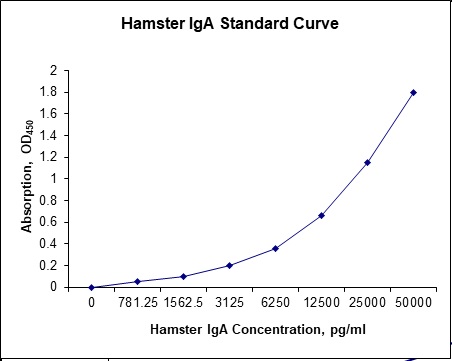
Standard Curve
Kit Components
1. Pre-coated 96-well Microplate
2. Biotinylated Detection Antibody
3. Streptavidin-HRP Conjugate
4. Lyophilized Standards
5. TMB One-Step Substrate
6. Stop Solution
7. 20 x PBS
8. Assay Buffer
Other Materials Required but not Provided:
1. Microplate Reader capable of measuring absorption at 450 nm
2. Log-log graph paper or computer and software for ELISA data analysis
3. Precision pipettes (1-1000 µl)
4. Multi-channel pipettes (300 µl)
5. Distilled or deionized water
Protocol Outline
1. Prepare all reagents, samples and standards as instructed in the datasheet.
2. Add 100 µl of Standard or samples to each well and incubate 1 h at RT.
3. Add 100 µl of Working Detection Antibody to each well and incubate 1 h at RT.
4. Add 100 µl of Working Streptavidin-HRP to each well and incubate 20 min at RT.
5. Add 100 µl of Substrate to each well and incubate 5-30 min at RT.
6. Add 50 µl of Stop Solution to each well and read at 450 nm immediately.
Background:
Immunoglobulin A (IgA) is an antibody that plays a critical role in mucosal immunity. More IgA is produced in mucosal linings than all other types of antibody combined;[1] between three and five grams are secreted into the intestinal lumen each day.[2] This accumulates to 75% of the total immunoglobulin produced in the entire body.[3] IgA has two subclasses (IgA1 and IgA2) and can exist in a dimeric form called secretory IgA (sIgA). In its secretory form, IgA is the main immunoglobulin found in mucous secretions, including tears, saliva, colostrum and secretions from the genitourinary tract, gastrointestinal tract, prostate and respiratory epithelium. It is also found in small amounts in blood. The secretory component of sIgA protects the immunoglobulin from being degraded by proteolytic enzymes, thus sIgA can survive in the harsh gastrointestinal tract environment and provide protection against microbes that multiply in body secretions. IgA is a poor activator of the complement system, and opsonises only weakly. Its heavy chains are of the type α. In the blood, IgA interacts with an Fc receptor called FcαRI (or CD89), which is expressed on immune effector cells, to initiate inflammatory reactions.[4] Ligation of FcαRI by IgA containing immune complexes causes antibody-dependent cell-mediated cytotoxicity (ADCC), degranulation of eosinophils and basophils, phagocytosis by monocytes, macrophages, and neutrophils, and triggering of respiratory burst activity by polymorphonuclear leukocytes.[4] Polymeric IgA (mainly the secretory dimer) is produced by plasma cells in the lamina propria adjacent to mucosal surfaces. It binds to the polymeric immunoglobulin receptor on the basolateral surface of epithelial cells, and is taken up into the cell via endocytosis. The receptor-IgA complex passes through the cellular compartments before being secreted on the luminal surface of the epithelial cells, still attached to the receptor. Proteolysis of the receptor occurs, and the dimeric IgA molecule, along with a portion of the receptor known as the secretory component, are free to diffuse throughout the lumen.[5] In the gut, it can bind to the mucus layer on top of the epithelial cells to form a barrier capable of neutralizing threats before they reach the cells. Decreased or absent IgA, termed selective IgA deficiency, can be a clinically significant immunodeficiency. Neisseria gonorrhœae (which causes gonorrhea), Streptococcus pneumoniae, and Haemophilus influenzae type B all releases a protease which destroys IgA.
Reference
- S Fagarasan and T Honjo (2003). Nat. Rev. Immunology 3 (1): 63–72.
- P. Brandtzaeg, R. Pabst (2004). Trends Immunology 25 (11): 570–577.
- AJ Macpherson and E Slack. (2007). Curr Opin Gastroenterol. 23 (6): 673–678.
- CS Kaetzel et al. (1991). Vet. Res. 37 (3): 455–67.
- CS Kaetzel et al. (1991) Proc Natl Acad Sci USA 88 (19): 8796–8800.

Product Citations
- Frutos J et al. (2018) Early feed restriction of lambs modifies ileal epimural microbiota and affects immunity parameters during the fattening period. Animal 12:10, 2115-2122. Impact factor: 1.654. Products used and cited: Nori Sheep IgA ELISA Kits (GR116001, GR116001-2)
- Wlazlo L et al. (2021) Effects of fermented rapseed meal on the gastrointestinal microbiota and immune status of rabbit. Animals 11:716. Impact factor: 2.62. Products used and cited: Nori Rabbit IgA, IgG and IgM ELISA Kits.
- Wlazlol, et al. (2021) Effect of fermented rapeseed meal on the gastrointestinal microbiota and immune status of rabbit (oryctolagus cuniculus). Animalsorg/10.3390/ani11030716. Impact factor: 1.654. Products used and cited: Nori Rabbit IgA, IgG and IgM ELISA Kits
- Martin A et al. (2022) Dietary Administration of L-Carnitine During the Fattening Period of Early Feed Restricted Lambs Modifies Ruminal Fermentation but Does Not Improve Feed Efficiency. frontiers in Physiol https://doi.org/10.3389/fphys.2022.840065. Impact factor: 4.0. Products used and cited: Nori Sheep IgA ELISA Kit.
Be the first to review “Nori Hamster IgA ELISA Kit”
You must be logged in to post a review.
Reviews
There are no reviews yet.