


Nori Equine GM-CSF ELISA Kit

Price range: $508.00 through $916.00
This ELISA kit is for quantification of GM-CSF in equine. This is a quick ELISA assay that reduces time to 50% compared to the conventional method, and the entire assay only takes 3 hours. This assay employs the quantitative sandwich enzyme immunoassay technique and uses biotin-streptavidin chemistry to improve the performance of the assays. An antibody specific for GM-CSF has been pre-coated onto a microplate. Standards and samples are pipetted into the wells and any GM-CSF present is bound by the immobilized antibody. After washing away any unbound substances, a detection antibody specific for GM-CSF is added to the wells. Following wash to remove any unbound antibody reagent, a detection reagent is added. After intensive wash a substrate solution is added to the wells and color develops in proportion to the amount of GM-CSF bound in the initial step. The color development is stopped, and the intensity of the color is measured.
Alternative names for GM-CSF: Granulocyte-macrophage colony-stimulating factor, CSF2
This product is for laboratory research use only, not for diagnostic and therapeutic purposes or any other purposes.
- Description
- How Elisa Works
- Documents
- Product Citations
- Reviews (0)
Description
Nori Equine GM-CSF ELISA Kit Summary
Alternative names for GM-CSF: Granulocyte-macrophage colony-stimulating factor, CSF2
Alternative names for equine: Horse
| Assay Type | Solid Phase Sandwich ELISA |
| Format | 96-well Microplate or 96-Well Strip Microplate |
| Method of Detection | Colorimetric |
| Number of Targets Detected | 1 |
| Target Antigen Accession Number | Q8WN17 |
| Assay Length | 3 hours |
| Quantitative/Semiquantitative | Quantitative |
| Sample Type | Plasma, Serum, Cell Culture, Urine, Cell/Tissue Lysates, Synovial Fluid, BAL, |
| Recommended Sample Dilution (Plasma/Serum) | No dilution for sample <ULOQ; sufficient dilution for samples >ULOQ |
| Sensitivity | 2 pg/mL |
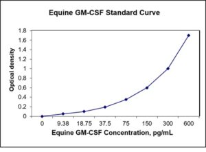
| Detection Range | 9.38-600 pg/mL |
| Specificity | Equine GM-CSF |
| Cross-Reactivity | < 0.5% cross-reactivity observed with available related molecules, < 50% cross-species reactivity observed with species tested. |
| Interference | No significant interference observed with available related molecules |
| Storage/Stability | 4 ºC for up to 6 months |
| Usage | For Laboratory Research Use Only. Not for diagnostic or therapeutic use. |
| Additional Notes | The kit allows for use in multiple experiments. |
Standard Curve
Kit Components
1. Pre-coated 96-well Microplate
2. Biotinylated Detection Antibody
3. Streptavidin-HRP Conjugate
4. Lyophilized Standards
5. TMB One-Step Substrate
6. Stop Solution
7. 20 x PBS
8. Assay Buffer
Other Materials Required but not Provided:
1. Microplate Reader capable of measuring absorption at 450 nm
2. Log-log graph paper or computer and software for ELISA data analysis
3. Precision pipettes (1-1000 µl)
4. Multi-channel pipettes (300 µl)
5. Distilled or deionized water
Protocol Outline
1. Prepare all reagents, samples and standards as instructed in the datasheet.
2. Add 100 µl of Standard or samples to each well and incubate 1 h at RT.
3. Add 100 µl of Working Detection Antibody to each well and incubate 1 h at RT.
4. Add 100 µl of Working Streptavidin-HRP to each well and incubate 20 min at RT.
5. Add 100 µl of Substrate to each well and incubate 5-30 min at RT.
6. Add 50 µl of Stop Solution to each well and read at 450 nm immediately.
Background:
Granulocyte-macrophage colony-stimulating factor (GM-CSF) is a protein secreted by macrophages, T cells, mast cells, NK cells, endothelial cells and fibroblasts (1, 2). GM-CSF is a cytokine that functions as a white blood cell growth factor and is also involved in tumor progression (3, 4). It is also a growth factor for erythroid, megakaryocyte, and eosinophil progenitors. On mature hematopoietic cells, GM-CSF is a survival factor and activates the effector functions of granulocytes, monocytes/macrophages, and eosinophils. GM-CSF promotes a Th1 biased immune response, angiogenesis, allergic inflammation, and the development of autoimmunity. GM-CSF stimulates stem cells to produce granulocytes (neutrophils, eosinophils, and basophils) and monocytes. Monocytes exit the circulation and migrate into tissue, whereupon they mature into macrophages and dendritic cells. Thus, it is part of the immune/inflammatory cascade, by which activation of a small number of macrophages can rapidly lead to an increase in their numbers, a process crucial for fighting infection. The active form of the protein is found extracellularly as a homodimer. The 22 kDa glycosylated GM-CSF, similar to IL-3 and IL-5, is a cytokine with a core of four bundled alpha helices. GM-CSF exerts its biological effects through a heterodimeric receptor complex composed of GM-CSF R alpha/CD116 and the signal transducing common beta chain (CD131) which is also a component of the high-affinity receptors for IL-3 and IL-5. In addition, GM-CSF binds a naturally occurring soluble form of GM-CSF R alpha. It shows clinical effectiveness in ameliorating chemotherapy-induced neutropenia, and GM-CSF transfected tumor cells are utilized as cancer vaccines. GM-CSF is found in high levels in joints with rheumatoid arthritis and blocking GM-CSF may reduce the inflammation or damage. Some drugs (e.g. MOR103) are being developed to block GM-CSF.
Reference
- Martinez-Moczygemba M, Huston DP (2003). J. Allergy Clin. Immunol. 112 (4): 653–65.
- Hamilton JA, Anderson GP (2005). Growth Factors 22 (4): 225–31.
- Mroczko B, Szmitkowski M (2005). Clin. Chem. Lab. Med. 42 (12): 1347–54.
- Morales JK, Kmieciak M, et al. (2009). Breast Cancer Res Treat. 123 (1): 39–49.

Be the first to review “Nori Equine GM-CSF ELISA Kit”
You must be logged in to post a review.
Reviews
There are no reviews yet.