


Nori Human ICAM-1 ELISA Kit

Price range: $508.00 through $916.00
This ELISA kit is for quantification of ICAM-1 in human. This is a quick ELISA assay that reduces time to 50% compared to the conventional method, and the entire assay only takes 3 hours. This assay employs the quantitative sandwich enzyme immunoassay technique and uses biotin-streptavidin chemistry to improve the performance of the assays. An antibody specific for ICAM-1 has been pre-coated onto a microplate. Standards and samples are pipetted into the wells and any ICAM-1 present is bound by the immobilized antibody. After washing away any unbound substances, a detection antibody specific for ICAM-1 is added to the wells. Following wash to remove any unbound antibody reagent, a detection reagent is added. After intensive wash a substrate solution is added to the wells and color develops in proportion to the amount of ICAM-1 bound in the initial step. The color development is stopped, and the intensity of the color is measured.
Alternative names for ICAM-1: Intercellular Adhesion Molecule 1, CD54
This product is for Laboratory Research Use Only not for diagnostic and therapeutic purposes or any other purposes.
- Description
- How Elisa Works
- Product Citations (1)
- Reviews (0)
Description
Nori Human ICAM-1 ELISA Kit Summary
Alternative names for ICAM-1: Intercellular Adhesion Molecule 1, CD54
| Assay Type | Solid Phase Sandwich ELISA |
| Format | 96-well Microplate or 96-Well Strip Microplate |
| Method of Detection | Colorimetric |
| Number of Targets Detected | 1 |
| Target Antigen Accession Number | P05363 |
| Assay Length | 3 hours |
| Quantitative/Semiquantitative | Quantitative |
| Sample Type | Plasma, Serum, Cell Culture, Urine, Cell/Tissue Lysates, Synovial Fluid, BAL, |
| Recommended Sample Dilution (Plasma/Serum) | No dilution for sample <ULOQ; sufficient dilution for samples >ULOQ |
| Sensitivity | 300 pg/mL |
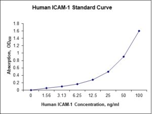
| Detection Range | 1.56-100 ng/mL |
| Specificity | Human ICAM-1 |
| Cross-Reactivity | < 0.5% cross-reactivity observed with available related molecules, < 50% cross-species reactivity observed with species tested. |
| Interference | No significant interference observed with available related molecules |
| Storage/Stability | 4 ºC for up to 6 months |
| Usage | For Laboratory Research Use Only. Not for diagnostic or therapeutic use. |
| Additional Notes | The kit allows for use in multiple experiments. |
Standard Curve
Kit Components
1. Pre-coated 96-well Microplate
2. Biotinylated Detection Antibody
3. Streptavidin-HRP Conjugate
4. Lyophilized Standards
5. TMB One-Step Substrate
6. Stop Solution
7. 20 x PBS
8. Assay Buffer
Other Materials Required but not Provided:
1. Microplate Reader capable of measuring absorption at 450 nm
2. Log-log graph paper or computer and software for ELISA data analysis
3. Precision pipettes (1-1000 µl)
4. Multi-channel pipettes (300 µl)
5. Distilled or deionized water
Protocol Outline
1. Prepare all reagents, samples and standards as instructed in the datasheet.
2. Add 100 µl of Standard or samples to each well and incubate 1 h at RT.
3. Add 100 µl of Working Detection Antibody to each well and incubate 1 h at RT.
4. Add 100 µl of Working Streptavidin-HRP to each well and incubate 20 min at RT.
5. Add 100 µl of Substrate to each well and incubate 5-30 min at RT.
6. Add 50 µl of Stop Solution to each well and read at 450 nm immediately.
Background:
ICAM-1 (Intercellular Adhesion Molecule 1) also known as CD54 is a protein that is encoded by the ICAM1 gene.[1] ICAM-1 is a member of the immunoglobulin superfamily and a transmembrane protein. ICAM-1 is a cell surface glycoprotein which is typically expressed on endothelial cells and cells of the immune system. It binds to integrins of type CD11a / CD18, or CD11b / CD18 and is also exploited by rhinovirus as a receptor for entry into respiratory epithelium.[2] ICAM-1 continuously present in low concentrations in the membranes of leukocytes and endothelial cells. Upon cytokine stimulation, the concentrations greatly increase. ICAM-1 can be induced by interleukin-1 and tumor necrosis factor and is expressed by the vascular endothelium, macrophages, and lymphocytes. ICAM-1 is a ligand for LFA-1 (integrin), a receptor found on leukocytes.[3] When activated, leukocytes bind to endothelial cells via ICAM-1/LFA-1 and then transmigrate into tissues.[4] LFA-1 has also been found in a soluble form,[5] which seems to bind and block ICAM-1.[6] ICAM-1 and soluble ICAM-1 have antagonistic effects on the tight junctions forming the blood-testis barrier, thus playing a major role in spermatogenesis.[7] ICAM-1 signaling seems to produce a recruitment of inflammatory immune cells such as macrophages and granulocytes.[8] ICAM-1 may also participate in a positive feedback loop and compete with ICAM-2 to maintain a proinflammatory environment conducive to leukocyte endothelial transmigration. ICAM-1 has been implicated in subarachnoid hemorrhage (SAH) and levels of ICAM-1 significantly elevated in patients.[9] ICAM-1 interacts with CD11a,[10] EZR[11] and CD18.[12]
References
- Carlson M, et al. (1988). Nucleic Acids Research. 16 (9): 4188. doi:1093/nar/16.9.4188.
- Bella J, et al. (1998). Proc Natl Acad Sci USA. 95 (8): 4140–45.
- Rothlein R, et al.. (1986). Journal of Immunology. 137 (4): 1270–4.
- Yang L, et al. (2005). Blood. 106 (2): 584–92. doi:1182/blood-2004-12-4942.
- Gjelstrup LC, et al. (2010). Journal of Immunology. 185 (7): 4154–68.
- Kragstrup TW, et al. (2014). Arthritis Research & Therapy. 16 (1): R42. doi:1186/ar4471.
- Xiao X, Mruk DD, Cheng CY (2013).Human Reproduction Update. 19 (2): 167–86.
- Etienne-Manneville S, et al. (1999). Journal of Immunology. 163 (2): 668–74.
- Polin RS, et al. (1998). Journal of Neurosurgery. 89 (4): 559–67.
- Shimaoka M, et al. (2003). Cell. 112 (1): 99–111.
- Heiska L, et al. (1998). The Journal of Biological Chemistry. 273 (34): 21893–900.
- Lu C, et al. (2001). The Journal of Biological Chemistry. 276 (18): 14642–48.

Product Citations
- Haranczyk M et al. (2022) Endothelial dysfunction in obstructive sleep apnea patients. Sleep and Breathing 26:231-242. org/10.1007/s11325-021-02382-4. Impact factor: 2.4. Products used and cited: Nori Human Endothelin-1, ICAM-1 ELISA Kit. Article
Be the first to review “Nori Human ICAM-1 ELISA Kit”
You must be logged in to post a review.
Reviews
There are no reviews yet.