


Nori Canine PDGF ELISA Kit

Price range: $461.00 through $832.00
This ELISA kit is for quantification of PDGF in canine. This is a quick ELISA assay that reduces time to 50% compared to the conventional method, and the entire assay only takes 3 hours. This assay employs the quantitative sandwich enzyme immunoassay technique and uses biotin-streptavidin chemistry to improve the performance of the assays. An antibody specific for PDGF has been pre-coated onto a microplate. Standards and samples are pipetted into the wells and any PDGF present is bound by the immobilized antibody. After washing away any unbound substances, a detection antibody specific for PDGF is added to the wells. Following wash to remove any unbound antibody reagent, a detection reagent is added. After intensive wash a substrate solution is added to the wells and color develops in proportion to the amount of PDGF bound in the initial step. The color development is stopped, and the intensity of the color is measured.
Alternative names for PDGF: Platelet-derived growth factor
This product is for Laboratory Research Use Only not for diagnostic and therapeutic purposes or any other purposes.
- Description
- How Elisa Works
- Product Citation (0)
- Reviews (0)
Description
Nori Canine PDGF ELISA Kit Summary
Alternative names for PDGF: Platelet-derived growth factor
Alternative names for canine: dog
| Assay Type | Solid Phase Sandwich ELISA |
| Format | 96-well Microplate or 96-Well Strip Microplate |
| Method of Detection | Colorimetric |
| Number of Targets Detected | 1 |
| Target Antigen Accession Number | NA |
| Assay Length | 3 hours |
| Quantitative/Semiquantitative | Quantitative |
| Sample Type | Plasma, Serum, Cell Culture, Urine, Cell/Tissue Lysates, Synovial Fluid, BAL, |
| Recommended Sample Dilution (Plasma/Serum) | No dilution for sample <ULOQ; sufficient dilution for samples >ULOQ |
| Sensitivity | 6 pg/mL |
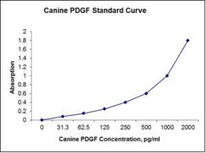
| Detection Range | 31.25-2000 pg/mL |
| Specificity | Canine PDGF |
| Cross-Reactivity | < 0.5% cross-reactivity observed with available related molecules, < 50% cross-species reactivity observed with species tested. |
| Interference | No significant interference observed with available related molecules |
| Storage/Stability | 4 ºC for up to 6 months |
| Usage | For Laboratory Research Use Only. Not for diagnostic or therapeutic use. |
| Additional Notes | The kit allows for use in multiple experiments. |
Standard Curve
Kit Components
1. Pre-coated 96-well Microplate
2. Biotinylated Detection Antibody
3. Streptavidin-HRP Conjugate
4. Lyophilized Standards
5. TMB One-Step Substrate
6. Stop Solution
7. 20 x PBS
8. Assay Buffer
Other Materials Required but not Provided:
1. Microplate Reader capable of measuring absorption at 450 nm
2. Log-log graph paper or computer and software for ELISA data analysis
3. Precision pipettes (1-1000 µl)
4. Multi-channel pipettes (300 µl)
5. Distilled or deionized water
Protocol Outline
1. Prepare all reagents, samples and standards as instructed in the datasheet.
2. Add 100 µl of Standard or samples to each well and incubate 1 h at RT.
3. Add 100 µl of Working Detection Antibody to each well and incubate 1 h at RT.
4. Add 100 µl of Working Streptavidin-HRP to each well and incubate 20 min at RT.
5. Add 100 µl of Substrate to each well and incubate 5-30 min at RT.
6. Add 50 µl of Stop Solution to each well and read at 450 nm immediately.
Background:
Platelet-derived growth factor (PDGF) is one of the numerous growth factors that regulate cell growth and division. In particular, it plays a significant role in blood vessel formation (angiogenesis), the growth of blood vessels from already-existing blood vessel tissue. Uncontrolled angiogenesis is a characteristic of cancer. PDGF is a dimeric glycoprotein composed of two A (-AA) or two B (-BB) chains or a combination of the two (-AB). PDGF[1][2] is a potent mitogen for cells of mesenchymal origin, including smooth muscle cells and glial cells. In both mouse and human, the PDGF signaling network consists of four ligands, PDGFA-D, and two receptors, PDGFR alpha and PDGFR beta. All PDGFs function as secreted, disulphide-linked homodimers, but only PDGFA and B can form functional heterodimers.
PDGF plays a role in embryonic development, cell proliferation, cell migration, and angiogenesis. PDGF has also been linked to several diseases such as atherosclerosis, fibrosis and malignant diseases. In addition, PDGF is a required element in cellular division for fibroblast. In essence, the PDGFs allow a cell to skip the G1 checkpoints in order to divide. PDGF and its receptors have provided a market for receptor antagonists to treat disease. Such antagonists include (but are not limited to) specific antibodies that target the molecule of interest, which act only in a neutralizing manner. Though it is synthesized[3] stored and released by platelets upon activation, it is produced by a plethora of cells including smooth muscle cells, activated macrophages, and endothelial cells[4]
References
- Hannink M, Donoghue DJ (1989). Biochim. Biophys. Acta 989 (1): 1–10.
- Heldin CH (1992). EMBO J. 11 (12): 4251–4259.
- Shulman T, et al. (July 1997). J. Biol. Chem. 272 (28): 17400–4.

Be the first to review “Nori Canine PDGF ELISA Kit”
You must be logged in to post a review.
Reviews
There are no reviews yet.