


Nori Canine SAA1 ELISA Kit

Price range: $508.00 through $916.00
This ELISA kit is for quantification of SAA1 in dog. This is a quick ELISA assay that reduces time to 50% compared to the conventional method, and the entire assay only takes 3 hours. This assay employs the quantitative sandwich enzyme immunoassay technique and uses biotin-streptavidin chemistry to improve the performance of the assays. An antibody specific for SAA1 has been pre-coated onto a microplate. Standards and samples are pipetted into the wells and any SAA1 present is bound by the immobilized antibody. After washing away any unbound substances, a detection antibody specific for SAA1 is added to the wells. Following wash to remove any unbound antibody reagent, a detection reagent is added. After intensive wash a substrate solution is added to the wells and color develops in proportion to the amount of SAA1 bound in the initial step. The color development is stopped, and the intensity of the color is measured.
Alternative names for SAA1: Serum amyloid A1
This product is for laboratory research use only not for diagnostic and therapeutic purposes or any other purposes.
- Description
- How Elisa Works
- Product Citations (2)
- Reviews (0)
Description
Nori Canine SAA1 ELISA Kit Summary
Alternative names for SAA1: Serum amyloid A1
Alternative name for canine: dog
| Assay Type | Solid Phase Sandwich ELISA |
| Format | 96-well Microplate or 96-Well Strip Microplate |
| Method of Detection | Colorimetric |
| Number of Targets Detected | 1 |
| Target Antigen Accession Number |
P19708 |
| Assay Length | 3 hours |
| Quantitative/Semiquantitative | Quantitative |
| Sample Type | Plasma, Serum, Cell Culture, Urine, Cell/Tissue Lysates, Synovial Fluid, BAL, |
| Recommended Sample Dilution (Plasma/Serum) | No dilution for sample <ULOQ; sufficient dilution for samples >ULOQ |
| Sensitivity | 250 pg/mL |
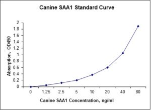
| Detection Range | 1.25-80 ng/mL |
| Specificity | Canine SAA1 |
| Cross-Reactivity | < 0.5% cross-reactivity observed with available related molecules, < 50% cross-species reactivity observed with species tested. |
| Interference | No significant interference observed with available related molecules |
| Storage/Stability | 4 ºC for up to 6 months |
| Usage | For Laboratory Research Use Only. Not for diagnostic or therapeutic use. |
| Additional Notes | The kit allows for use in multiple experiments. |
Standard Curve
Kit Components
1. Pre-coated 96-well Microplate
2. Biotinylated Detection Antibody
3. Streptavidin-HRP Conjugate
4. Lyophilized Standards
5. TMB One-Step Substrate
6. Stop Solution
7. 20 x PBS
8. Assay Buffer
Other Materials Required but not Provided:
1. Microplate Reader capable of measuring absorption at 450 nm
2. Log-log graph paper or computer and software for ELISA data analysis
3. Precision pipettes (1-1000 µl)
4. Multi-channel pipettes (300 µl)
5. Distilled or deionized water
Protocol Outline
1. Prepare all reagents, samples and standards as instructed in the datasheet.
2. Add 100 µl of Standard or samples to each well and incubate 1 h at RT.
3. Add 100 µl of Working Detection Antibody to each well and incubate 1 h at RT.
4. Add 100 µl of Working Streptavidin-HRP to each well and incubate 20 min at RT.
5. Add 100 µl of Substrate to each well and incubate 5-30 min at RT.
6. Add 50 µl of Stop Solution to each well and read at 450 nm immediately.
Background:
Serum amyloid A (SAA) proteins are a family of apolipoproteins associated with high-density lipoprotein (HDL) in plasma. Different isoforms of SAA are expressed constitutively (constitutive SAAs) at different levels or in response to inflammatory stimuli (acute phase SAAs). These proteins are produced predominantly by the liver.[1] Acute-phase SAA proteins (A-SAAs) are secreted during the acute phase of inflammation. These proteins have several roles, including the transport of cholesterol to the liver for secretion into the bile, the recruitment of immune cells to inflammatory sites, and the induction of enzymes that degrade extracellular matrix. A-SAAs are implicated in several chronic inflammatory diseases, such as amyloidosis, atherosclerosis, and rheumatoid arthritis.[2] Three acute-phase SAA isoforms have been reported in mice, called SAA1, SAA2, and SAA3. During inflammation, SAA1 and SAA2 are expressed and induced principally in the liver, whereas SAA3 is induced in many distinct tissues. SAA1 and SAA2 genes are regulated in liver cells by the proinflammatory cytokines IL-1, IL-6, and TNF-α. Both SAA1 and SAA2 are induced up to a 1000-fold in mice under acute inflammatory conditions following exposure to bacterial lipopolysaccharide (LPS).[2] SAA is also an acute phase marker. Similar to CRP, levels of acute-phase SAA increase within hours after inflammatory stimulus, and the magnitude of increase may be greater than that of CRP. Relatively trivial inflammatory stimuli can lead to SAA responses. It has been suggested that SAA levels correlate better with disease activity in early inflammatory joint disease than do ESR and CRP. Although largely produced by hepatocytes, SAA is produced by adipocytes as well, and its serum concentration is associated with body mass index. A fourth SAA (SAA4) was identified in Sheeps and is expressed constitutively in the liver and, thus, is defined as a constitutive SAA (C-SAA).[4] The originally designated SAA5 in the Sheep is now called SAA4.[5][6]
References
- Uhlar CM, Whitehead AS (1999). Eur. J. Biochem.265 (2): 501–23.
- Zhang N, et al. (2005). J. Immunol.174 (12): 8125–34.
- Betts JC, et al. (1991). Scand. J. Immunol. 34 (4): 471–82.
- Steel DM, et al. (1993). Genomics 16 (2): 447–54.
- de Beer MC, et al. (1994). J. Biol. Chem. 269 (6): 4661–7.
- de Beer MC, et al. (1996). Genomics 34(1): 139–42.

Product Citations
- Farag VMEM et al. (2017) Serum amyloid A4 and ceruloplasmain evaluated mastitic cattle with Escherichia coli or staphylococcus aureus including resistant genes. J Bioanalysis & Biomed 9:132-136. Doi:10.4172/1948-593x.1000167. Impact factor: 2.80. Product used and cited: Nori Bovine SAA4 ELISA Kit, GR112079.
- El-Ashker M et al. (2014) Gentamicin-induced acute kidney injury in equines is associated with marked acute phase response: an experimental study on donkey (Equus asinus). J Vet Sci Med Diagn 4:2. Impact factor: 0.65. Products used and cited: Nori Equine IL-1β, IL-6, IFN-g, IL-10, SAA, Haptoglobin, CRP and SA ELISA kits.
Be the first to review “Nori Canine SAA1 ELISA Kit”
You must be logged in to post a review.

Reviews
There are no reviews yet.