


Nori Canine HIP-1 ELISA Kit

Price range: $508.00 through $916.00
This ELISA kit is for quantification of HIP-1 in canine. This is a quick ELISA assay that reduces time to 50% compared to the conventional method, and the entire assay only takes 3 hours. This assay employs the quantitative sandwich enzyme immunoassay technique and uses biotin-streptavidin chemistry to improve the performance of the assays. An antibody specific for HIP1 has been pre-coated onto a microplate. Standards and samples are pipetted into the wells and any HIP1 present is bound by the immobilized antibody. After washing away any unbound substances, a detection antibody specific for HIP1 is added to the wells. Following wash to remove any unbound antibody reagent, a detection reagent is added. After intensive wash a substrate solution is added to the wells and color develops in proportion to the amount of HIP1 bound in the initial step. The color development is stopped, and the intensity of the color is measured.
Alternative names for HIP-1: HIP1, Huntingtin-interacting protein 1
This product is for laboratory research use only not for diagnostic and therapeutic purposes or any other purposes.
- Description
- How Elisa Works
- Documents
- Product Citation (0)
- Reviews (0)
Description
Nori Canine HIP-1 ELISA Kit Summary
Alternative names for HIP-1, HIP1, Huntingtin-interacting protein 1
Alternative names for canine: Dog
| Assay Type | Solid Phase Sandwich ELISA |
| Format | 96-well Microplate or 96-Well Strip Microplate |
| Method of Detection | Colorimetric |
| Number of Targets Detected | 1 |
| Target Antigen Accession Number | A0A8I3MQX2 |
| Assay Length | 3 hours |
| Quantitative/Semiquantitative | Quantitative |
| Sample Type | Plasma, Serum, Cell Culture, Urine, Cell/Tissue Lysates, Synovial Fluid, BAL, |
| Recommended Sample Dilution (Plasma/Serum) | No dilution for sample <ULOQ; sufficient dilution for samples >ULOQ |
| Sensitivity | 60 pg/mL |
| Detection Range | 0.313-20 ng/mL |
| Specificity | Canine HIP-1 |
| Cross-Reactivity | < 0.5% cross-reactivity observed with available related molecules, < 50% cross-species reactivity observed with species tested. |
| Interference | No significant interference observed with available related molecules |
| Storage/Stability | 4 ºC for up to 6 months |
| Usage | For Laboratory Research Use Only. Not for diagnostic or therapeutic use. |
| Additional Notes | The kit allows for use in multiple experiments. |
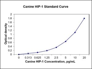
Standard Curve
Kit Components
1. Pre-coated 96-well Microplate
2. Biotinylated Detection Antibody
3. Streptavidin-HRP Conjugate
4. Lyophilized Standards
5. TMB One-Step Substrate
6. Stop Solution
7. 20 x PBS
8. Assay Buffer
Other Materials Required but not Provided:
1. Microplate Reader capable of measuring absorption at 450 nm
2. Log-log graph paper or computer and software for ELISA data analysis
3. Precision pipettes (1-1000 µl)
4. Multi-channel pipettes (300 µl)
5. Distilled or deionized water
Protocol Outline
1. Prepare all reagents, samples and standards as instructed in the datasheet.
2. Add 100 µl of Standard or samples to each well and incubate 1 h at RT.
3. Add 100 µl of Working Detection Antibody to each well and incubate 1 h at RT.
4. Add 100 µl of Working Streptavidin-HRP to each well and incubate 20 min at RT.
5. Add 100 µl of Substrate to each well and incubate 5-30 min at RT.
6. Add 50 µl of Stop Solution to each well and read at 450 nm immediately.
Background:
Huntingtin-interacting protein 1 (HIP-1) is a protein that is encoded by the HIP1 gene.[1]
Hip-1 interacts with the huntingtin protein. HIP-1 contains a domain homologous to the death effector domains. It is believed that accumulation of high levels of the free form of HIP-1 in the cell is one of the mechanisms by which neuron cell death is caused in Huntington’s disease (via the caspase-3 route). The role of Hip-1 in caspase mediated cell death remains unclear. HIP1 was found to bind to Htt in an N-terminal dependent manner, and co-localizes with Htt in the CNS although the nature of this interaction with respect to muHtt was not identified. The CAG expansion seen with muHtt results in decreased binding affinity for HIP1, thus causing disruption of HIP1’s usual function, and also an increase in free HIP1.[2] It is likely that this decreased affinity plays a role in mediating HD pathogenesis, due to loss of cytoskeletal integrity and induction of apoptosis. HIP1’s pro apoptotic effect may involve activation of caspase-8 and a novel HIP1 protein interactor HIPPI.[3] HIP1’s non-pathological activity includes clathrin assembly via interaction with clathrin light chains.[4] HIP1 is the human homologue of Sla2p, a membrane protein in the periphery.[5] Actin binding by Hip-1 is altered depending on whether clathrin is also bound to Hip-1. HIP1 has been found to be overexpressed in some cancers including a subset of colorectal and prostate cancers.[6] HIP1 can regulate transcription of hormone receptors via the androgen response element (ARE) and alter the rate of degradation of the AR.[7] HIP1 may regulate, or at least interact with proteins that also possess the ARE.
References
- Wanker EE, et al. (1997). Human Molecular Genetics. 6(3): 487–95.
- Hackam AS, et al. (2000). The Journal of Biological Chemistry. 275(52): 41299–308.
- Gervais FG, et al. (2002). Nature Cell Biology. 4(2): 95–105.
- Legendre-Guillemin V, et al. (2005). Journal of Biological Chemistry. 280(7): 6101–8.
- Holzmann C, et al. (2001). Molecular Brain Research. 92(1–2): 85–97.
- Rao DS, et al. (2002). The Journal of Clinical Investigation. 110(3): 351–60.
- Mills IG, et al. (2005). The Journal of Cell Biology. 170(2): 191–200.

Be the first to review “Nori Canine HIP-1 ELISA Kit”
You must be logged in to post a review.

Reviews
There are no reviews yet.