


Nori Rat Galectin-1 ELISA Kit

Price range: $508.00 through $916.00
This ELISA kit is for quantification of galectin-1 in rat. This is a quick ELISA assay that reduces time to 50% compared to the conventional method, and the entire assay only takes 3 hours. This assay employs the quantitative sandwich enzyme immunoassay technique and uses biotin-streptavidin chemistry to improve the performance of the assays. An antibody specific for galectin-1 has been pre-coated onto a microplate. Standards and samples are pipetted into the wells and any galectin-1 present is bound by the immobilized antibody. After washing away any unbound substances, a detection antibody specific for galectin-1 is added to the wells. Following wash to remove any unbound antibody reagent, a detection reagent is added. After intensive wash a substrate solution is added to the wells and color develops in proportion to the amount of galectin-1 bound in the initial step. The color development is stopped, and the intensity of the color is measured.
Alternative names for galectin-1: LGALS1
This product is for laboratory research use only not for diagnostic and therapeutic purposes or any other purposes.
- Description
- How Elisa Works
- Documents
- Product Citations
- Reviews (0)
Description
Nori Rat Galectin-1 ELISA Kit Summary
Alternative names for galectin-1: LGALS1
| Assay Type | Solid Phase Sandwich ELISA |
| Format | 96-well Microplate or 96-Well Strip Microplate |
| Method of Detection | Colorimetric |
| Number of Targets Detected | 1 |
| Target Antigen Accession Number |
P11762 |
| Assay Length | 3 hours |
| Quantitative/Semiquantitative | Quantitative |
| Sample Type | Plasma, Serum, Cell Culture, Urine, Cell/Tissue Lysates, Synovial Fluid, BAL, |
| Recommended Sample Dilution (Plasma/Serum) | No dilution for sample <ULOQ; sufficient dilution for samples >ULOQ |
| Sensitivity | 60 pg/mL |
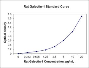
| Detection Range | 0.313-20 ng/mL |
| Specificity | Rat Galectin-1 |
| Cross-Reactivity | < 0.5% cross-reactivity observed with available related molecules, < 50% cross-species reactivity observed with species tested. |
| Interference | No significant interference observed with available related molecules |
| Storage/Stability | 4 ºC for up to 6 months |
| Usage | For Laboratory Research Use Only. Not for diagnostic or therapeutic use. |
| Additional Notes | The kit allows for use in multiple experiments. |
Standard Curve
Kit Components
1. Pre-coated 96-well Microplate
2. Biotinylated Detection Antibody
3. Streptavidin-HRP Conjugate
4. Lyophilized Standards
5. TMB One-Step Substrate
6. Stop Solution
7. 20 x PBS
8. Assay Buffer
Other Materials Required but not Provided:
1. Microplate Reader capable of measuring absorption at 450 nm
2. Log-log graph paper or computer and software for ELISA data analysis
3. Precision pipettes (1-1000 µl)
4. Multi-channel pipettes (300 µl)
5. Distilled or deionized water
Protocol Outline
1. Prepare all reagents, samples and standards as instructed in the datasheet.
2. Add 100 µl of Standard or samples to each well and incubate 1 h at RT.
3. Add 100 µl of Working Detection Antibody to each well and incubate 1 h at RT.
4. Add 100 µl of Working Streptavidin-HRP to each well and incubate 20 min at RT.
5. Add 100 µl of Substrate to each well and incubate 5-30 min at RT.
6. Add 50 µl of Stop Solution to each well and read at 450 nm immediately.
Background:
Galectin-1 is a protein that is encoded by the LGALS1 gene.[1] The galectins are a family of beta-galactoside-binding proteins implicated in modulating cell-cell and cell-matrix interactions.[2] Galectin-1 may act as an autocrine negative growth factor that regulates cell proliferation. Galectin-1 expression in Hodgkin Lymphoma has also been shown to mediate immunosuppression of CD8+ T-cells.[3] Galectin-1 binds oncogenic H-Ras to mediate Ras membrane anchorage and cell transformation.[4] Galectin-1 induces the differentiation of Dendritic cells towards a phenotype which dampens T helper 1 cells and T helper 17 cells and dampens inflammation via interleukin-10 and interleukin-27.[5] It also plays a role in the formation and expression of HLA-G in the syncytium.[6] Galectin-1 is expressed by the endometrial stromal cells throughout the menstrual cycle, however significantly increases during implantation. It can be found in the nucleus, the cytoplasm, the cell surface and in the extracellular space. Galectins in general lack a traditional signal sequence, but are still secreted across the plasma membrane. This non-traditional secretion requires a functional glycan binding site. Galectin 1 contains a single carbohydrate recognition domain through which it can bind glycans both as a monomer and as a homodimer. Dimers are non-covalently bound and will spontaneously disassociate in low concentration.[7] Galectin 1 does not bind glycans when oxidized.[8] Having 6 cysteine residues, the oxidation state has a significant effect on the protein structure. The oxidized form is reported to have alternative functions not involving carbohydrate binding.[9]
References
- Gauthier L, et al. (2002). Proc Natl Acad Sci U S A. 99 (20): 13014–9.
- Gandhi, MK (2007). Blood. 110 (4): 1326–9.
- Paz A, et al. (2001). Oncogene. 20 (51): 7486–93.
- Ilarregui JM, et al. (2009). Nat. Immunol. 10 (9): 981–91.
- Comninos AN, et al. (2014). Hum. Reprod. Update. 20 (2): 153–74.
- Cho M, Cummings RD (1995). J. Biol. Chem. 270 (10): 5198–206.
- Outenreath RL, Jones AL (1992). J. Neurocytol. 21 (11): 788–95.
- Kadoya T, Horie H (2005). Curr Drug Targets. 6 (4): 375–83.

Be the first to review “Nori Rat Galectin-1 ELISA Kit”
You must be logged in to post a review.
Reviews
There are no reviews yet.