


Nori Rat Amphiregulin (AREG) ELISA Kit

Price range: $508.00 through $916.00
This ELISA kit is for quantification of AREG in rat. This is a quick ELISA assay that reduces time to 50% compared to the conventional method, and the entire assay only takes 3 hours. This assay employs the quantitative sandwich enzyme immunoassay technique and uses biotin-streptavidin chemistry to improve the performance of the assays. An antibody specific for AREG has been pre-coated onto a microplate. Standards and samples are pipetted into the wells and any AREG present is bound by the immobilized antibody. After washing away any unbound substances, a detection antibody specific for AREG is added to the wells. Following wash to remove any unbound antibody reagent, a detection reagent is added. After intensive wash a substrate solution is added to the wells and color develops in proportion to the amount of AREG bound in the initial step. The color development is stopped, and the intensity of the color is measured.
Alternative names for AREG: Amphiregulin
This product is for Laboratory Research Use Only not for diagnostic and therapeutic purposes or any other purposes.
- Description
- How Elisa Works
- Product Citation (0)
- Reviews (0)
Description
Nori Rat AREG ELISA Kit Summary
Alternative names for AREG: Amphiregulin, SDGF
| Assay Type | Solid Phase Sandwich ELISA |
| Format | 96-well Microplate or 96-Well Strip Microplate |
| Method of Detection | Colorimetric |
| Number of Targets Detected | 1 |
| Target Antigen Accession Number | P24338 |
| Assay Length | 3 hours |
| Quantitative/Semiquantitative | Quantitative |
| Sample Type | Plasma, Serum, Cell Culture, Urine, Cell/Tissue Lysates, Synovial Fluid, BAL, |
| Recommended Sample Dilution (Plasma/Serum) | No dilution for sample <ULOQ; sufficient dilution for samples >ULOQ |
| Sensitivity | 6 pg/mL |
| Detection Range | 31.25-2000 pg/mL |
| Specificity | Rat AREG |
| Cross-Reactivity | < 0.5% cross-reactivity observed with available related molecules, < 50% cross-species reactivity observed with species tested. |
| Interference | No significant interference observed with available related molecules |
| Storage/Stability | 4 ºC for up to 6 months |
| Usage | For Laboratory Research Use Only. Not for diagnostic or therapeutic use. |
| Additional Notes | The kit allows for use in multiple experiments. |
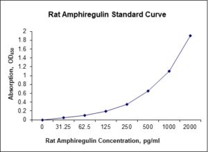
Standard Curve
Kit Components
1. Pre-coated 96-well Microplate
2. Biotinylated Detection Antibody
3. Streptavidin-HRP Conjugate
4. Lyophilized Standards
5. TMB One-Step Substrate
6. Stop Solution
7. 20 x PBS
8. Assay Buffer
Other Materials Required but not Provided:
1. Microplate Reader capable of measuring absorption at 450 nm
2. Log-log graph paper or computer and software for ELISA data analysis
3. Precision pipettes (1-1000 µl)
4. Multi-channel pipettes (300 µl)
5. Distilled or deionized water
Protocol Outline
1. Prepare all reagents, samples and standards as instructed in the datasheet.
2. Add 100 µl of Standard or samples to each well and incubate 1 h at RT.
3. Add 100 µl of Working Detection Antibody to each well and incubate 1 h at RT.
4. Add 100 µl of Working Streptavidin-HRP to each well and incubate 20 min at RT.
5. Add 100 µl of Substrate to each well and incubate 5-30 min at RT.
6. Add 50 µl of Stop Solution to each well and read at 450 nm immediately.
Background:
Amphiregulin (AREG) is a transmembrane glycoprotein with 252 amino acids that is encoded by the AREG gene.[1][2] AREG is a member of the epidermal growth factor (EGF) family and a critical autocrine growth factor as well as a mitogen for astrocytes, Schwann cells, and fibroblasts. It is a ligand for EGF and related to transforming growth factor alpha (TGF-alpha). AREG interacts with the EGF receptor (EGFR) to promote the growth of normal epithelial cells. AREG is a critical factor in estrogen action and ductal development of the mammary glands.[3][4] Amphiregulin is essential for mammary ductal development, as evidenced by absence of ductal growth in amphiregulin knockout mice,[4] a similar phenotypes of EGFR and ERα knockout mice.[4] Amphiregulin is expressed in many tissues and can be induced by TGF-α, TNF-α, interleukin 1, and prostaglandins.[5] Overexpression of amphiregulin is connected with various cancer.[5] Expression of AREG is connected with proliferation of fibroblasts and production of proinflammatory cytokines interleukin 8 and vascular endothelial growth factor (VEGF).[6] Gene mutations are associated with a psoriasis-like skin phenotype.[16] Higher circulating levels of amphiregulin are associated with AGVHD progression.[7] Amphiregulin is part of cellular response type 2.[8] It was found that the cell source of amphiregulin is innate lymphoid cells 2 (ILC2) which are dependent on interleukin 33. ILC2 expressed amphiregulin after tissue damage of the intestines and activation by IL-33. Moreover, endogenous AREG with IL-33 decreased the intestinal inflammation in mice with normal count of T-lymphocytes and in deficient mice.[9]
References
- Shoyab M, et al. (1989). Science. 243 (4894 Pt 1): 1074–6.
- Plowman GD, et al. (1990). Molecular and Cellular Biology. 10 (5): 1969–81.
- Aupperlee MD, et al. (2013). Breast Cancer Research. 15 (3): R44. doi:1186/bcr3431.
- McBryan J, et al. (2008). Journal of Mammary Gland Biology and Neoplasia. 13 (2): 159–69.
- Busser B, et al. (2011). Biochimica et Biophysica Acta- Reviews on Cancer. 1816 (2): 119–31.
- Yamane S, et al. (2008). Journal of Inflammation. 5: 5.
- Bhagavathula N, et al. (2005). The American Journal of Pathology. 166 (4): 1009–16.
- Zaiss DM, et al. (2006). Science. 314 (5806): 1746.
- Monticelli LA, et al. (2015). Proc Natl Acad Sci USA. 112 (34): 10762–7.

Be the first to review “Nori Rat Amphiregulin (AREG) ELISA Kit”
You must be logged in to post a review.
Reviews
There are no reviews yet.