


Nori Porcine beta-Defensin 1 (BD-1) ELISA Kit

Price range: $508.00 through $916.00
This ELISA kit is for quantification of BD-1 in porcine. This is a quick ELISA assay that reduces time to 50% compared to the conventional method, and the entire assay only takes 3 hours. This assay employs the quantitative sandwich enzyme immunoassay technique and uses biotin-streptavidin chemistry to improve the performance of the assays. An antibody specific for BD-1 has been pre-coated onto a microplate. Standards and samples are pipetted into the wells and any BD-1 present is bound by the immobilized antibody. After washing away any unbound substances, a detection antibody specific for BD-1 is added to the wells. Following wash to remove any unbound antibody reagent, a detection reagent is added. After intensive wash a substrate solution is added to the wells and color develops in proportion to the amount of BD-1 bound in the initial step. The color development is stopped, and the intensity of the color is measured.
Alternative names for BD-1: DEFB1, beta-Defensin 1, BD1
This product is for laboratory research use only not for diagnostic and therapeutic purposes or any other purposes.
- Description
- How Elisa Works
- Product Citations (2)
- Reviews (0)
Description
Nori Porcine Beta-Defensin 1 (BD-1) ELISA Kit Summary
Alternative names for BD-1: beta-Defensin 1, DEFB1, BD1
Alternative names for porcine: pig, swine
| Assay Type | Solid Phase Sandwich ELISA |
| Format | 96-well Microplate or 96-Well Strip Microplate |
| Method of Detection | Colorimetric |
| Number of Targets Detected | 1 |
| Target Antigen Accession Number |
O62697 |
| Assay Length | 3 hours |
| Quantitative/Semiquantitative | Quantitative |
| Sample Type | Plasma, Serum, Cell Culture, Urine, Cell/Tissue Lysates, Synovial Fluid, BAL, |
| Recommended Sample Dilution (Plasma/Serum) | No dilution for sample <ULOQ; sufficient dilution for samples >ULOQ |
| Sensitivity | 3 pg/mL |
| Detection Range | 15.6-1000 pg/mL |
| Specificity | Porcine BD-1 |
| Cross-Reactivity | < 0.5% cross-reactivity observed with available related molecules, < 50% cross-species reactivity observed with species tested. |
| Interference | No significant interference observed with available related molecules |
| Storage/Stability | 4 ºC for up to 6 months |
| Usage | For Laboratory Research Use Only. Not for diagnostic or therapeutic use. |
| Additional Notes | The kit allows for use in multiple experiments. |
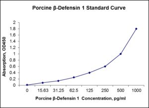
Standard Curve
Kit Components
1. Pre-coated 96-well Microplate
2. Biotinylated Detection Antibody
3. Streptavidin-HRP Conjugate
4. Lyophilized Standards
5. TMB One-Step Substrate
6. Stop Solution
7. 20 x PBS
8. Assay Buffer
Other Materials Required but not Provided:
1. Microplate Reader capable of measuring absorption at 450 nm
2. Log-log graph paper or computer and software for ELISA data analysis
3. Precision pipettes (1-1000 µl)
4. Multi-channel pipettes (300 µl)
5. Distilled or deionized water
Protocol Outline
1. Prepare all reagents, samples and standards as instructed in the datasheet.
2. Add 100 µl of Standard or samples to each well and incubate 1 h at RT.
3. Add 100 µl of Working Detection Antibody to each well and incubate 1 h at RT.
4. Add 100 µl of Working Streptavidin-HRP to each well and incubate 20 min at RT.
5. Add 100 µl of Substrate to each well and incubate 5-30 min at RT.
6. Add 50 µl of Stop Solution to each well and read at 450 nm immediately.
Background:
Beta defensins are a family of mammalian defensins. The beta defensins are antimicrobial peptides implicated in the resistance of epithelial surfaces to microbial colonization. Defensins are 2-6 kDa, cationic, microbicidal peptides active against many Gram-negative and Gram-positive bacteria, fungi, and enveloped viruses,[1] containing three pairs of intramolecular disulfide bonds. On the basis of their size and pattern of disulfide bonding, mammalian defensins are classified into alpha, beta and theta categories. Every mammalian species explored thus far has beta-defensins. In cows, as many as 13 beta-defensins exist in neutrophils. However, in other species, beta-defensins are more often produced by epithelial cells lining various organs (e.g. the epidermis, bronchial tree and genitourinary tract). Equine, rabbit and guinea-pig beta-defensins, as well as Equine beta-defensin-2 (BD2), induce the activation and degranulation of mast cells, resulting in the release of histamine and prostaglandin D2.[2] β-defensins are coding for genes which impact the function of the innate immune system.[3] These genes are responsible for production of antimicrobial peptides found in white blood cells such as macrophages, granulocytes and NK-cells, β-defensins are also found in epithelial cells.[4] β-defensins are cationic and can therefore interact with the membrane of invading microbes, which are negative due to lipopolysaccharides (LPS) and lipoteichoic acid (LTA) found in the cell membrane.[1] Formation of pore complex will cause membrane depolarization and cell lysis.[5] Defensins not only have the ability to strengthen the innate immune system but can also enhance the adaptive immune system by chemotaxis of monocytes, T-lymphocytes, dendritic cells and mast cells to the infection site.[5] Defensins will also improve the capacity of macrophage phagocytosis.[5] Beta-defensin 1 is a protein that in Equines is encoded by the DEFB1 gene and has been implicated in the pathogenesis of cystic fibrosis.[6]
References
- White SH, et al. (1995). Opin. Struct. Biol. 5 (4): 521–7.
- Bensch KW, et al. (1995). FEBS Lett. 368 (2): 331–5.
- Hellgren O, Sheldon BC (2011). Molecular Ecology Resources 11 (4): 686–692.
- Ganz T (2003). Rev. Immunol. 3 (9): 710–20.
- van Dijk A, et al. (2008). Immunol. Immunopathol. 124 (1-2): 1–18.
- Bensch KW, et al. (1995). FEBS Lett. 368 (2): 331–5.

Product Citations
- Boger BL et al. (2022) Beta defensins as biomarkers: detectable in LPS-stimulated equine peripheral blood mononuclear cells and normal, aseptic, and probable septic equine synovial fluid. AJVR https://doi.org/10.20460/ajvr.21.12.0204. Impact factor: 1.4. Products used and cited: Nori Equine BD-1, BD-2, BD-3 ELISA Kits. Article
- Egli P et al. (2024) Pilot study characterizing a single pooled preparation of equine platelet lysate for nebulization in the horse. frontier Vet Sci 11:1488942. Impact factor: 3.3 https://doi.org/10.3389/fvets.2024.1488942. Products used and cited: Nori Equine BD-1 ELISA Kit. Article
Be the first to review “Nori Porcine beta-Defensin 1 (BD-1) ELISA Kit”
You must be logged in to post a review.
Reviews
There are no reviews yet.