




















Nori Mouse S100A1 ELISA Kit
Price range: $508.00 through $916.00
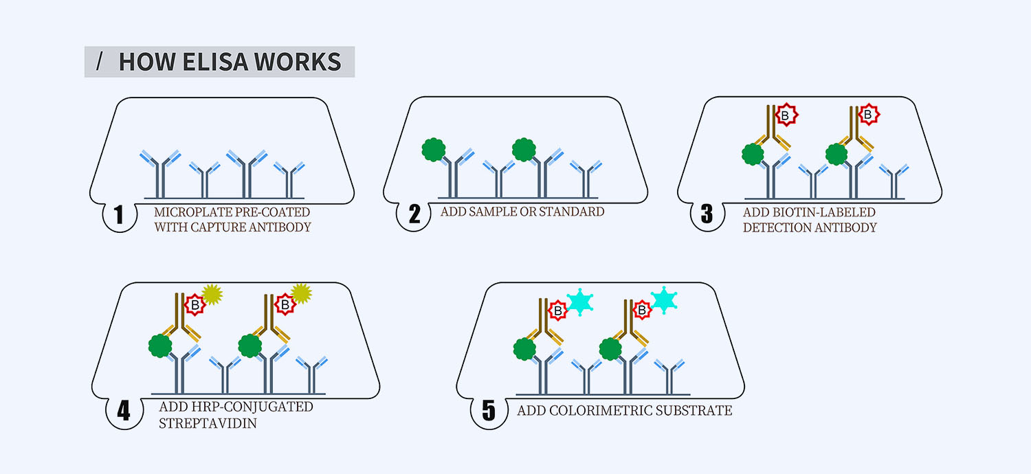
This ELISA kit is for quantification of S100A1 in mouse. This is a quick ELISA assay that reduces time to 50% compared to the conventional method, and the entire assay only takes 3 hours. This assay employs the quantitative sandwich enzyme immunoassay technique and uses biotin-streptavidin chemistry to improve the performance of the assays. An antibody specific for S100A1 has been pre-coated onto a microplate. Standards and samples are pipetted into the wells and any S100A1 present is bound by the immobilized antibody. After washing away any unbound substances, a detection antibody specific for S100A1 is added to the wells. Following wash to remove any unbound antibody reagent, a detection reagent is added. After intensive wash a substrate solution is added to the wells and color develops in proportion to the amount of S100A1 bound in the initial step. The color development is stopped, and the intensity of the color is measured.
Alternative names for S100A1: S100 calcium-binding protein A1
This product is for laboratory research use only not for diagnostic and therapeutic purposes or any other purposes.
- Description
- How Elisa Works
- Documents
- Product Citation (0)
- Reviews (0)
Description
Nori Mouse S100A1 ELISA Kit Summary
Alternative names for S100A1: S100 calcium-binding protein A1
| Assay Type | Solid Phase Sandwich ELISA |
| Format | 96-well Microplate or 96-Well Strip Microplate |
| Method of Detection | Colorimetric |
| Number of Targets Detected | 1 |
| Target Antigen Accession Number | P56565 |
| Assay Length | 3 hours |
| Quantitative/Semiquantitative | Quantitative |
| Sample Type | Plasma, Serum, Cell Culture, Urine, Cell/Tissue Lysates, Synovial Fluid, BAL, |
| Recommended Sample Dilution (Plasma/Serum) | No dilution for sample <ULOQ; sufficient dilution for samples >ULOQ |
| Sensitivity | 30 pg/mL |
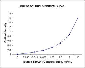
| Detection Range | 0.156-10 ng/mL |
| Specificity | Mouse S100A1 |
| Cross-Reactivity | < 0.5% cross-reactivity observed with available related molecules, < 50% cross-species reactivity observed with species tested. |
| Interference | No significant interference observed with available related molecules |
| Storage/Stability | 4 ºC for up to 6 months |
| Usage | For Laboratory Research Use Only. Not for diagnostic or therapeutic use. |
| Additional Notes | The kit allows for use in multiple experiments. |
Standard Curve
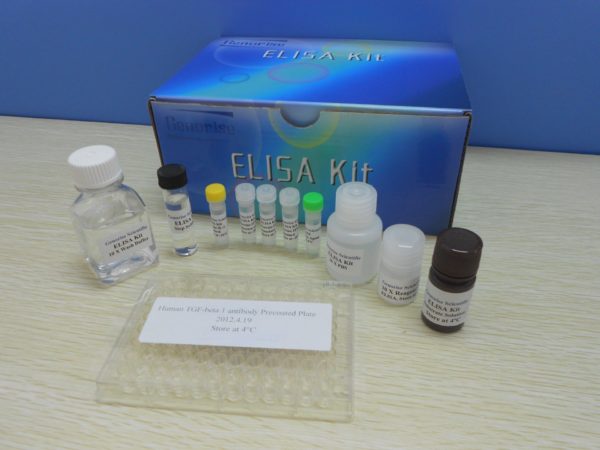
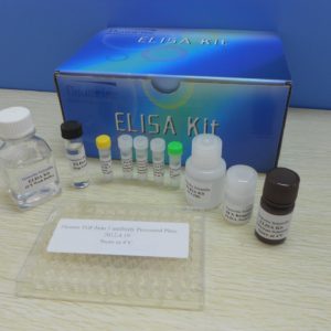
Kit Components
1. Pre-coated 96-well Microplate
2. Biotinylated Detection Antibody
3. Streptavidin-HRP Conjugate
4. Lyophilized Standards
5. TMB One-Step Substrate
6. Stop Solution
7. 20 x PBS
8. Assay Buffer
Other Materials Required but not Provided:
1. Microplate Reader capable of measuring absorption at 450 nm
2. Log-log graph paper or computer and software for ELISA data analysis
3. Precision pipettes (1-1000 µl)
4. Multi-channel pipettes (300 µl)
5. Distilled or deionized water
Protocol Outline
1. Prepare all reagents, samples and standards as instructed in the datasheet.
2. Add 100 µl of Standard or samples to each well and incubate 1 h at RT.
3. Add 100 µl of Working Detection Antibody to each well and incubate 1 h at RT.
4. Add 100 µl of Working Streptavidin-HRP to each well and incubate 20 min at RT.
5. Add 100 µl of Substrate to each well and incubate 5-30 min at RT.
6. Add 50 µl of Stop Solution to each well and read at 450 nm immediately.
Background:
S100A1, also known as S100 calcium-binding protein A1 is a protein which is encoded by the S100A1 gene.[1] S100A1 is highly expressed in cardiac and skeletal muscle, and localizes to Z-discs and sarcoplasmic reticulum. S100A1 has shown promise as an effective candidate for gene therapy to treat post-myocardially infarcted cardiac tissue. S100 proteins are localized in the cytoplasm and/or nucleus of a wide range of cells, and involved in the regulation of a number of cellular processes such as cell cycle progression and differentiation. This protein may function in stimulation of Ca2+-induced Ca2+ release, inhibition of microtubule assembly, and inhibition of protein kinase C-mediated phosphorylation. S100A1 is expressed during development in the primitive heart at embryonic day 8 in levels that are similar between atria and ventricles. As development progresses up to embryonic day 17.5, S100A1 expression shifts to a lower levels in atria and higher levels in ventricular myocardium.[2] S100A1 has shown to be a regulator of myocardial contractility. In animal models of disease, S100A1 protein levels has been shown to be altered in right ventricular hypertrophied tissue in a model of pulmonary hypertension;[3] several tissue types (brain, skeletal muscle and cardiac muscle) in a model of type I diabetes mellitus;[4] S100A1 has been demonstrated as a regulator of the genetic program underlying cardiac hypertrophy, in that S100A1 inhibits alpha1 adrenergic stimulation of hypertrophic genes, including MYH7, ACTA1 and S100B.[5] S100A1 has also been identified as a novel regulator of endothelial cell post-ischemic angiogenesis, as patients with limb ischemia exhibited downregulation of S100A1 expression in hypoxic tissue.[6]
References
- Morii K, et al. (1991). Biochem Biophysics Research Communications. 175 (1): 185–91.
- Kiewitz R, et al. (2000). Biochimica et Biophysica Acta. 1498 (2–3): 207–19.
- Ehlermann P, et al. (2000). Biochimica et Biophysica Acta. 1500 (2): 249–55.
- Zimmer DB, et al. (1997). Endocrinology. 138 (12): 5176–83.
- Tsoporis JN, et al. (2003). Molecular and Cellular Biochemistry. 242 (1–2): 27–33.
- Descamps, B; et al. (2013). Circulation Research. 112 (1): 3–5.
Be the first to review “Nori Mouse S100A1 ELISA Kit”
You must be logged in to post a review.
Reviews
There are no reviews yet.