





















Nori Mouse PKC alpha ELISA Kit
Price range: $508.00 through $916.00
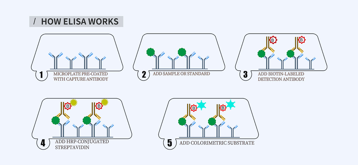
This ELISA kit is for quantification of PKCA in mouse. This is a quick ELISA assay that reduces time to 50% compared to the conventional method, and the entire assay only takes 3 hours. This assay employs the quantitative sandwich enzyme immunoassay technique and uses biotin-streptavidin chemistry to improve the performance of the assays. An antibody specific for PKCA has been pre-coated onto a microplate. Standards and samples are pipetted into the wells and any PKCA present is bound by the immobilized antibody. After washing away any unbound substances, a detection antibody specific for PKCA is added to the wells. Following wash to remove any unbound antibody reagent, a detection reagent is added. After intensive wash a substrate solution is added to the wells and color develops in proportion to the amount of PKCA bound in the initial step. The color development is stopped, and the intensity of the color is measured.
Alternative names for PKC alpha: Protein kinase C alpha, PKCA, PRKCA
This product is for laboratory research use only not for diagnostic and therapeutic purposes or any other purposes.
- Description
- How Elisa Works
- Documents
- Product Citation (0)
- Reviews (0)
Description
Nori Mouse PKC alpha ELISA Kit Summary
Alternative names for PKC alpha: Protein kinase C alpha, PKCA, PRKCA
| Assay Type | Solid Phase Sandwich ELISA |
| Format | 96-well Microplate or 96-Well Strip Microplate |
| Method of Detection | Colorimetric |
| Number of Targets Detected | 1 |
| Target Antigen Accession Number | P20444 |
| Assay Length | 3 hours |
| Quantitative/Semiquantitative | Quantitative |
| Sample Type | Plasma, Serum, Cell Culture, Urine, Cell/Tissue Lysates, Synovial Fluid, BAL, |
| Recommended Sample Dilution (Plasma/Serum) | No dilution for sample <ULOQ; sufficient dilution for samples >ULOQ |
| Sensitivity | 30 pg/mL |
| Detection Range | 0.156-10 ng/mL |
| Specificity | Mouse PKCA |
| Cross-Reactivity | < 0.5% cross-reactivity observed with available related molecules, < 50% cross-species reactivity observed with species tested. |
| Interference | No significant interference observed with available related molecules |
| Storage/Stability | 4 ºC for up to 6 months |
| Usage | For Laboratory Research Use Only. Not for diagnostic or therapeutic use. |
| Additional Notes | The kit allows for use in multiple experiments. |
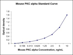
Standard Curve
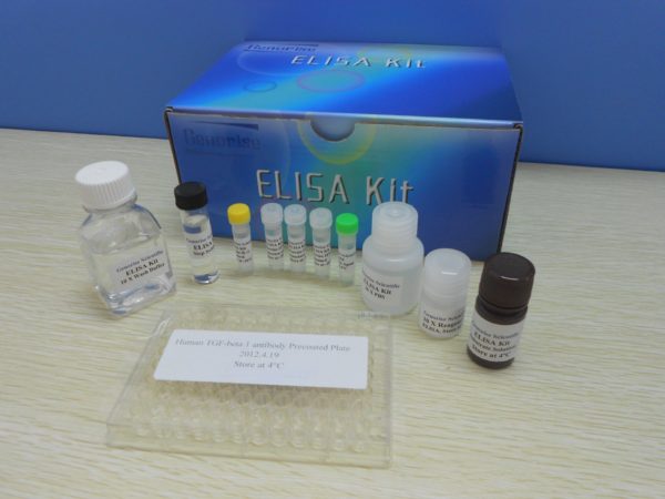
Kit Components
1. Pre-coated 96-well Microplate
2. Biotinylated Detection Antibody
3. Streptavidin-HRP Conjugate
4. Lyophilized Standards
5. TMB One-Step Substrate
6. Stop Solution
7. 20 x PBS
8. Assay Buffer
Other Materials Required but not Provided:
1. Microplate Reader capable of measuring absorption at 450 nm
2. Log-log graph paper or computer and software for ELISA data analysis
3. Precision pipettes (1-1000 µl)
4. Multi-channel pipettes (300 µl)
5. Distilled or deionized water
Protocol Outline
1. Prepare all reagents, samples and standards as instructed in the datasheet.
2. Add 100 µl of Standard or samples to each well and incubate 1 h at RT.
3. Add 100 µl of Working Detection Antibody to each well and incubate 1 h at RT.
4. Add 100 µl of Working Streptavidin-HRP to each well and incubate 20 min at RT.
5. Add 100 µl of Substrate to each well and incubate 5-30 min at RT.
6. Add 50 µl of Stop Solution to each well and read at 450 nm immediately.
Background:
Protein kinase C alpha (PKCα) is an enzyme that is encoded by the PRKCA gene and is a member of PKC family. PKCα plays roles in many different cellular processes, such as cell adhesion, cell transformation, cell cycle checkpoint, and cell volume control. PKCα may be a fundamental regulator of cardiac contractility and Ca2+ handling in myocytes. PKC-α is unique in its mode of regulation compared to other kinases within this family. The primary mode of PKC-α’s regulation involves its interaction with the cell membrane, not direct interaction with specific molecules.[1] At warmer temperatures, phospholipids exist in a more fluid state as a result of increased intramolecular motion. The more fluid the cell membrane, the greater PKC-α’s activity. Phospholipids that solidify at an irregular or angled orientation with respect to the membrane, can reduce PKC-α’s activity.[1] The composition of the cell membrane can also affect PKC-α’s function. Membranes with long hydrophobic domains result in decreased activity because it is harder for PKC-α to insert into the membrane. At low concentrations, the hydrophobic domain is shorter allowing PKC-α to readily insert into the membrane and its activity increases.[1] PKC-α plays a vital role in epithelial tissue and is involved in altering the function of tight junctions. Cells infected with certain types of epithelial cancer exhibit increased PKC-α activity. This is a result of a change in the shape of the cell membrane, particularly in the areas where tight junctions exists.[2] With greater activity of PKC-α, the tight junctions lose their ability to form a tight barrier.[3] Increased activation of PKCα is associated with the growth and invasion of cancers.[4] High levels of PKCα are linked to malignant brain cancer.[5] Moreover, a high proliferation rate of glioma tumor cells is the result of overexpression of isozyme PKCα.[6] PKC-α shows important regulation of phospholipase D and point mutations at particular phenylalanine residues have shown to inhibit PKC-α’s ability to activate phospholipase D.[7] PKC-α is correlated with the differentiation of erythroid progenitor cells in bone marrow.[8]
References
- Micol V, et al. (1999). Biophysical Journal. 76 (2): 916–27.
- Mullin JM, et al. (2000). Annals of the New York Academy of Sciences. 915: 231–6.
- Rosson D, et al. (1997). The Journal of Biological Chemistry. 272(23): 14950–3.
- Haughian JM, Bradford AP (2009). Journal of Cellular Physiology. 220 (1): 112–8.
- Yazaki T, et al. (1996). Molecular Pharmacology. 50 (2): 236–42.
- Baltuch GH, et al. (1995). Journal of Neuro-Oncology. 24 (3): 241–50.
- Hu T, Exton JH (2005). Biochem Biophysic Res Communications. 333 (3): 750–3.
- Myklebust JH, et al. (2000). Blood. 95 (2): 510–8.
Be the first to review “Nori Mouse PKC alpha ELISA Kit”
You must be logged in to post a review.
Reviews
There are no reviews yet.