




















Nori Human PD1 ELISA Kit
Price range: $508.00 through $916.00
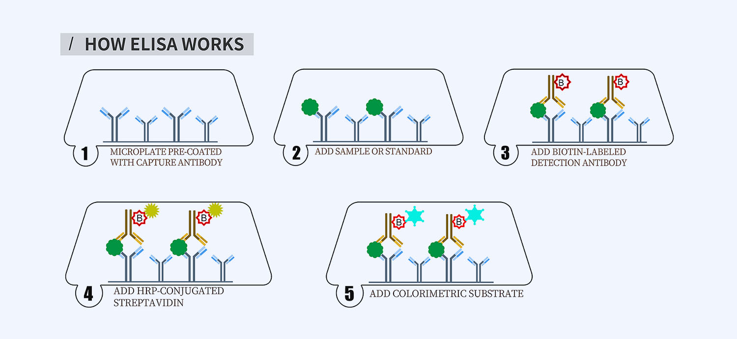
This ELISA kit is for quantification of PD1 in human. This is a quick ELISA assay that reduces time to 50% compared to the conventional method, and the entire assay only takes 3 hours. This assay employs the quantitative sandwich enzyme immunoassay technique and uses biotin-streptavidin chemistry to improve the performance of the assays. An antibody specific for PD1 has been pre-coated onto a microplate. Standards and samples are pipetted into the wells and any PD1 present is bound by the immobilized antibody. After washing away any unbound substances, a detection antibody specific for PD1 is added to the wells. Following wash to remove any unbound antibody reagent, a detection reagent is added. After intensive wash a substrate solution is added to the wells and color develops in proportion to the amount of PD1 bound in the initial step. The color development is stopped, and the intensity of the color is measured.
Alternative names for PD1: Programmed cell death protein 1, PD-1, CD279, cluster of differentiation 279, PCD1, PDCD1
This product is for Laboratory Research Use Only not for diagnostic and therapeutic purposes or any other purposes.
- Description
- How Elisa Works
- Documents
- Product Citations (0)
- Reviews (0)
Description
Nori Human PD1 ELISA Kit Summary
Alternative names for PD1: Programmed cell death protein 1, PD-1, CD279, cluster of differentiation 279, PCD1, PDCD1
| Assay Type | Solid Phase Sandwich ELISA |
| Format | 96-well Microplate or 96-Well Strip Microplate |
| Method of Detection | Colorimetric |
| Number of Targets Detected | 1 |
| Target Antigen Accession Number | Q15116 |
| Assay Length | 3 hours |
| Quantitative/Semiquantitative | Quantitative |
| Sample Type | Plasma, Serum, Cell Culture, Urine, Cell/Tissue Lysates, Synovial Fluid, BAL, |
| Recommended Sample Dilution (Plasma/Serum) | No dilution for sample <ULOQ; sufficient dilution for samples >ULOQ |
| Sensitivity | 6 pg/mL |
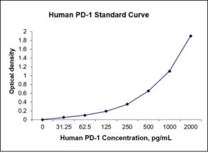
| Detection Range | 31.25-2000 pg/mL |
| Specificity | Human PD1 |
| Cross-Reactivity | < 0.5% cross-reactivity observed with available related molecules, < 50% cross-species reactivity observed with species tested. |
| Interference | No significant interference observed with available related molecules |
| Storage/Stability | 4 ºC for up to 6 months |
| Usage | For Laboratory Research Use Only. Not for diagnostic or therapeutic use. |
| Additional Notes | The kit allows for use in multiple experiments. |
Standard Curve
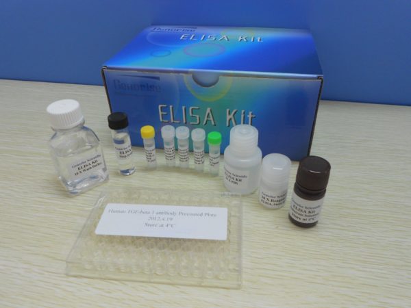
Kit Components
1. Pre-coated 96-well Microplate
2. Biotinylated Detection Antibody
3. Streptavidin-HRP Conjugate
4. Lyophilized Standards
5. TMB One-Step Substrate
6. Stop Solution
7. 20 x PBS
8. Assay Buffer
Other Materials Required but not Provided:
1. Microplate Reader capable of measuring absorption at 450 nm
2. Log-log graph paper or computer and software for ELISA data analysis
3. Precision pipettes (1-1000 µl)
4. Multi-channel pipettes (300 µl)
5. Distilled or deionized water
Protocol Outline
1. Prepare all reagents, samples and standards as instructed in the datasheet.
2. Add 100 µl of Standard or samples to each well and incubate 1 h at RT.
3. Add 100 µl of Working Detection Antibody to each well and incubate 1 h at RT.
4. Add 100 µl of Working Streptavidin-HRP to each well and incubate 20 min at RT.
5. Add 100 µl of Substrate to each well and incubate 5-30 min at RT.
6. Add 50 µl of Stop Solution to each well and read at 450 nm immediately.
Background:
Programmed cell death protein 1 ( PD-1) also known as CD279 (cluster of differentiation 279) encoded by PCD1 gene,[1] is a protein on the surface of T and B cells that has a role in regulating the immune system‘s response to the cells by down-regulating the immune system and promoting self-tolerance by suppressing T cell inflammatory activity. This prevents autoimmune diseases, but it can also prevent the immune system from killing cancer cells.[2] PD-1 is an immune checkpoint and guards against autoimmunity through two mechanisms. First, it promotes apoptosis of antigen-specific T-cells in lymph nodes. Second, it reduces apoptosis in regulatory T cells.[3] The PD-1 is a cell surface receptor that binds two ligands, PD-L1 and PD-L2. PD-L1 is upregulated on macrophages and dendritic cells (DC) in response to LPS and GM-CSF treatment.[4] PD-1 and its ligands negatively regulate immune responses. PD-1 knockout mice have been shown to develop lupus-like glomerulonephritis and dilated cardiomyopathy on the C57BL/6 and BALB/c backgrounds, respectively.[5] Blocking of PD-1 leads to a reduction in cerebral amyloid-β plaques and improves cognitive performance in mice.[6] Immune blockade of PD-1 evoked an IFN-γ dependent immune response that recruited monocyte-derived macrophages to the brain that were then capable of clearing the amyloid-β plaques from the tissue. Expression of PD-L1 on tumor cells inhibits anti-tumor activity through engagement of PD-1 on effector T cells.[7] Triggering PD-1, expressed on monocytes and up-regulated upon monocytes activation, by its ligand PD-L1 induces IL-10 production which inhibits CD4 T-cell function.[8] Overexpression of PD1 on CD8+ T cells is one of the indicators of T-cell exhaustion (e.g. in chronic infection or cancer).[2][9]
References
- Ishida Y, et al. (1992). The EMBO Journal. 11 (11): 3887–95.
- Francisco LM, et al. (2010). Immunological Reviews. 236: 219–42.
- Fife BT, Pauken KE (2011). Annals of the New York Academy of Sciences. 1217 (1): 45–59.
- Iwai Yet al. (2002). Proc Natl Acad Sci USA. 99 (19): 12293–7.
- Nishimura H, et al. (2001). Science. 291 (5502): 319–22.
- Kurnellas MP, et al. (2013). Science Translational Medicine. 5 (179): 179ra42.
- Nishimura H, et al. (1999). Immunity. 11 (2): 141–51.
- Pauken KE, Wherry EJ (2015). Trends in Immunology. 36 (4): 265–76.
- Wang X, et al. (2016). OncoTargets and Therapy. 9: 5023–39.
Product Citations
Be the first to review “Nori Human PD1 ELISA Kit”
You must be logged in to post a review.
Reviews
There are no reviews yet.