


Nori Human CD39 ELISA Kit

Price range: $508.00 through $916.00
This ELISA kit is for quantification of CD39 in human. This is a quick ELISA assay that reduces time to 50% compared to the conventional method, and the entire assay only takes 3 hours. This assay employs the quantitative sandwich enzyme immunoassay technique and uses biotin-streptavidin chemistry to improve the performance of the assays. An antibody specific for CD39 has been pre-coated onto a microplate. Standards and samples are pipetted into the wells and any CD39 present is bound by the immobilized antibody. After washing away any unbound substances, a detection antibody specific for CD39 is added to the wells. Following wash to remove any unbound antibody reagent, a detection reagent is added. After intensive wash a substrate solution is added to the wells and color develops in proportion to the amount of CD39 bound in the initial step. The color development is stopped, and the intensity of the color is measured.
Alternative names for CD39: Cluster of Differentiation 39, diphosphohydrolase-1 (gene: ENTPD1; protein: NTPDase1)
This product is for Laboratory Research Use Only not for diagnostic and therapeutic purposes or any other purposes.
- Description
- How Elisa Works
- Product Citation (0)
- Reviews (0)
Description
Nori Human CD39 ELISA Kit Summary
Alternative names for CD39: Cluster of Differentiation 39, diphosphohydrolase-1 (gene: ENTPD1; protein: NTPDase1)
| Assay Type | Solid Phase Sandwich ELISA |
| Format | 96-well Microplate or 96-Well Strip Microplate |
| Method of Detection | Colorimetric |
| Number of Targets Detected | 1 |
| Target Antigen Accession Number | P49951 |
| Assay Length | 3 hours |
| Quantitative/Semiquantitative | Quantitative |
| Sample Type | Plasma, Serum, Cell Culture, Urine, Cell/Tissue Lysates, Synovial Fluid, BAL, |
| Recommended Sample Dilution (Plasma/Serum) | No dilution for sample <ULOQ; sufficient dilution for samples >ULOQ |
| Sensitivity | 5 pg/mL |
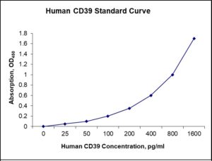
| Detection Range | 25-1600 pg/mL |
| Specificity | Natural and recombinant human CD39 |
| Cross-Reactivity | < 0.5% cross-reactivity observed with available related molecules, < 50% cross-species reactivity observed with species tested. |
| Interference | No significant interference observed with available related molecules |
| Storage/Stability | 4 ºC for up to 6 months |
| Usage | For Laboratory Research Use Only. Not for diagnostic or therapeutic use. |
| Additional Notes | The kit allows for use in multiple experiments. |
Standard Curve
Kit Components
1. Pre-coated 96-well Microplate
2. Biotinylated Detection Antibody
3. Streptavidin-HRP Conjugate
4. Lyophilized Standards
5. TMB One-Step Substrate
6. Stop Solution
7. 20 x PBS
8. Assay Buffer
Other Materials Required but not Provided:
1. Microplate Reader capable of measuring absorption at 450 nm
2. Log-log graph paper or computer and software for ELISA data analysis
3. Precision pipettes (1-1000 µl)
4. Multi-channel pipettes (300 µl)
5. Distilled or deionized water
Protocol Outline
1. Prepare all reagents, samples and standards as instructed in the datasheet.
2. Add 100 µl of Standard or samples to each well and incubate 1 h at RT.
3. Add 100 µl of Working Detection Antibody to each well and incubate 1 h at RT.
4. Add 100 µl of Working Streptavidin-HRP to each well and incubate 20 min at RT.
5. Add 100 µl of Substrate to each well and incubate 5-30 min at RT.
6. Add 50 µl of Stop Solution to each well and read at 450 nm immediately.
Background:
Ectonucleoside triphosphate diphosphohydrolase-1 (gene: ENTPD1; protein: NTPDase1) also known as CD39 (Cluster of Differentiation 39), is a typical cell surface-located enzymes with an extracellularly facing catalytic site.[1] NTPDase1 is an ectonucleotidase that catalyse the hydrolysis of γ- and β-phosphate residues of triphospho- and diphospho-nucleosides to the monophospho-nucleoside derivative.[2] For example, NTPDase1 hydrolyzes P2 receptor ligands, namely ATP, ADP, UTP and UDP with similar efficacy.[3] NTPDase1 can therefore affect P2 receptor activation and functions.[4] Oxidized low-density lipoproteins enhance expression and activity of CD39 and CD73 in the human aortic valve endothelium.[5] T-cell expression of CD39 was higher in acute exacerbations of chronic obstructive pulmonary disease patients than stable COPD patients or healthy controls.[6] Ablation of CD73 minimally effects in vivo thrombosis, but increased CD39 expression on hematopoietic-derived cells,especially monocytes, attenuates in vivo arterial thrombosis.[7] Th17(CD39+) cells are markedly diminished and fail to generate AMP/adenosine, thereby limiting control of both target cell proliferation and IL-17 production in juvenile autoimmune liver disease (AILD).[8]
References
- Kaczmarek E, et al. (1996). The Journal of Biological Chemistry. 271 (51): 33116–22.
- Yegutkin GG (2008). Biochimica et Biophysica Acta. 1783 (5): 673–94.
- Kukulski Fet al. (2005). Purinergic Signalling. 1 (2): 193–204.
- Kukulski F, et al. (2011). Advances in Pharmacology. 61: 263–99.
- Kaniewska-Bednarczuk E, et al. (2016) Nucleosides Nucleotides Nucleic Acids 35 (10-12), 713-719.
- Tan DB et al. (2016) Hum. Immunol. 77 (10), 916-920.
- Covarrubias R, et al. (2016) Arterioscler. Thromb. Vasc. Biol. 36 (9), 1809-1820.
- Liberal R, et al. (2016) J. Autoimmun. 72, 102-112.

Be the first to review “Nori Human CD39 ELISA Kit”
You must be logged in to post a review.
Reviews
There are no reviews yet.