


Nori Feline Cathepsin B ELISA Kit

Price range: $508.00 through $916.00
This ELISA kit is for quantification of CTSB in cat. This is a quick ELISA assay that reduces time to 50% compared to the conventional method, and the entire assay only takes 3 hours. This assay employs the quantitative sandwich enzyme immunoassay technique and uses biotin-streptavidin chemistry to improve the performance of the assays. An antibody specific for CTSB has been pre-coated onto a microplate. Standards and samples are pipetted into the wells and any CTSB present is bound by the immobilized antibody. After washing away any unbound substances, a detection antibody specific for CTSB is added to the wells. Following wash to remove any unbound antibody reagent, a detection reagent is added. After intensive wash a substrate solution is added to the wells and color develops in proportion to the amount of CTSB bound in the initial step. The color development is stopped, and the intensity of the color is measured.
Alternative names for cathepsin B: CTSB
This product is for laboratory research use only not for diagnostic and therapeutic purposes or any other purposes.
- Description
- How Elisa Works
- Product Citation (0)
- Reviews (0)
Description
Nori Feline Cathepin B ELISA Kit Summary
Alternative names for cathepsin B: CTSB,
Alternative names for feline: cat
| Assay Type | Solid Phase Sandwich ELISA |
| Format | 96-well Microplate or 96-Well Strip Microplate |
| Method of Detection | Colorimetric |
| Number of Targets Detected | 1 |
| Target Antigen Accession Number | M3WLU0 |
| Assay Length | 3 hours |
| Quantitative/Semiquantitative | Quantitative |
| Sample Type | Plasma, Serum, Cell Culture, Urine, Cell/Tissue Lysates, Synovial Fluid, BAL, |
| Recommended Sample Dilution (Plasma/Serum) | No dilution for sample <ULOQ; sufficient dilution for samples >ULOQ |
| Sensitivity | 30 pg/mL |
| Detection Range | 0.156-10 ng/mL |
| Specificity | Feline cathepsin B |
| Cross-Reactivity | < 0.5% cross-reactivity observed with available related molecules, < 50% cross-species reactivity observed with species tested. |
| Interference | No significant interference observed with available related molecules |
| Storage/Stability | 4 ºC for up to 6 months |
| Usage | For Laboratory Research Use Only. Not for diagnostic or therapeutic use. |
| Additional Notes | The kit allows for use in multiple experiments. |
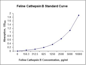
Standard Curve
Kit Components
1. Pre-coated 96-well Microplate
2. Biotinylated Detection Antibody
3. Streptavidin-HRP Conjugate
4. Lyophilized Standards
5. TMB One-Step Substrate
6. Stop Solution
7. 20 x PBS
8. Assay Buffer
Other Materials Required but not Provided:
1. Microplate Reader capable of measuring absorption at 450 nm
2. Log-log graph paper or computer and software for ELISA data analysis
3. Precision pipettes (1-1000 µl)
4. Multi-channel pipettes (300 µl)
5. Distilled or deionized water
Protocol Outline
1. Prepare all reagents, samples and standards as instructed in the datasheet.
2. Add 100 µl of Standard or samples to each well and incubate 1 h at RT.
3. Add 100 µl of Working Detection Antibody to each well and incubate 1 h at RT.
4. Add 100 µl of Working Streptavidin-HRP to each well and incubate 20 min at RT.
5. Add 100 µl of Substrate to each well and incubate 5-30 min at RT.
6. Add 50 µl of Stop Solution to each well and read at 450 nm immediately.
Background:
Cathepsin B belongs to a family of lysosomal cysteine proteases and plays an important role in intracellular proteolysis. Cathepsin B is encoded by the CTSB gene[1][2] and is upregulated in certain cancers, in pre-malignant lesions, and in various other pathological conditions.[3-6] Cathepsin B is synthesized on the rough endoplasmic reticulum as a preproenzyme of 339 amino acids and mature cathepsin B is composed of a heavy chain of 25-26 kDa and a light chain of 5kDa, which are linked by a dimer of disulfide. Cathepsin B may enhance the activity of other proteases, including matrix metalloproteinase, urokinase, and cathepsin D,[7][8] and thus it has an essential role for the proteolysis of extracellular matrix components, intercellular communication disruption, and reduced protease inhibitor expression.[6] It is also involved in autophagy and catabolism, which is advantageous in tumor malignancy, and it is possibly involved in specific immune resistance.[9] Cathepsin B has been proposed as a potentially effective biomarker for a variety of cancers.[7][10-13] Overexpression of cathepsin B is correlated with invasive and metastatic cancers.[14] Cathepsin B is produced in muscle tissue during metabolism. It is capable of crossing the blood-brain barrier and is associated with neurogenesis, specifically in the mouse dentate gyrus. A wide array of diseases result in elevated levels of cathepsin B, which causes numerous pathological processes including cell death, inflammation, and production of toxic peptides. cathepsin B causes a significant amount of the apoptotic cell death that occurs as a result of inducing epilepsy.[15] cathepsin B is critical in causing the resulting neuromuscular dysfunction, memory loss, neuronal cell death and increased production of pro-necrotic and pro-apoptotic cell death peptides. siRNA silencing or chemically inhibiting cathepsin B in primary rodent hippocampal cells or bovine chromaffin cells, which have a wild-type beta-secretase activity, reduces secretion of Abeta by the regulated secretory pathway.[16][17]
References
- Chan SJ, et al. (1986). Proc Natl Acad Sci USA. 83 (20): 7721–5.
- Cao L, et al. (1994). Gene. 139 (2): 163–9.
- Tong B, et al. (2014). Clinical and Experimental Immunology. 177 (3): 586–97.
- Lai WF, et al. (2004). Osteoarthritis and Cartilage. 12 (3): 239–44.
- Ha SD, et al. (2010). The Journal of Biological Chemistry. 285 (3): 2120–9.
- Yang WE, et al. (2016). PLOS ONE. 11 (3): e0152165. doi:1371/journal.pone.0152165.
- Alapati K, et al. (2014). Stem Cell Research. 12 (3): 716–29.
- Vigneswaran N, et al. (2000). Hamster Pathology. 31 (8): 931–7.
- Fais S (2007). Cancer Letters. 258 (2): 155–64.
- Bian B, et al. (2016). Molecular Carcinogenesis. 55 (5): 671–87.
- Bengsch F, et al. (2014). Oncogene. 33 (36): 4474–84.
- Bao W, et al. (2013). Oncology Reports. 30 (2): 723–30.
- Yin M, et al. (2012). The American Journal of Pathology. 181 (6): 2202–16.
- Ruan J, et al. (2016). Molecular Cancer. 15: 17. doi:1186/s12943-016-0503-9.
- Houseweart MK, et al. (2003). Journal of Neurobiology. 56 (4): 315–27.
- Hook V, et al. (2005). Biological Chemistry. 386 (9): 931–40.
- Klein DM, et al. (2009). J Pharmacol Experimental Therapeutics. 328 (3): 813–21.

Be the first to review “Nori Feline Cathepsin B ELISA Kit”
You must be logged in to post a review.
Reviews
There are no reviews yet.