




















Nori Equine KL-6 (MUC1) ELISA Kit
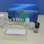
Price range: $508.00 through $916.00
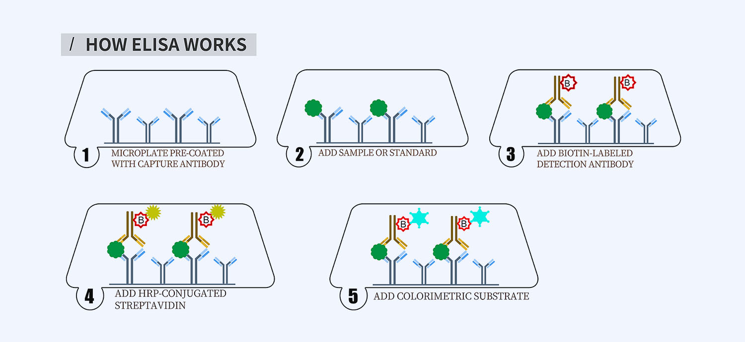
This ELISA kit is for quantification of KL-6 (MUC1) in equine. This is a quick ELISA assay that reduces time to 50% compared to the conventional method, and the entire assay only takes 3 hours. This assay employs the quantitative sandwich enzyme immunoassay technique and uses biotin-streptavidin chemistry to improve the performance of the assays. An antibody specific for KL-6 has been pre-coated onto a microplate. Standards and samples are pipetted into the wells and any KL-6 present is bound by the immobilized antibody. After washing away any unbound substances, a detection antibody specific for KL-6 is added to the wells. Following wash to remove any unbound antibody reagent, a detection reagent is added. After intensive wash a substrate solution is added to the wells and color develops in proportion to the amount of KL-6 bound in the initial step. The color development is stopped, and the intensity of the color is measured.
Alternative names for KL-6 (MUC1): Mucin 1, polymorphic epithelial mucin, PEM, epithelial membrane antigen, EMA
This product is for laboratory research use only not for diagnostic and therapeutic purposes or any other purposes.
- Description
- How Elisa Works
- Documents
- Product Citation (0)
- Reviews (0)
Description
Nori Equine KL-6 (MUC1) ELISA Kit Summary
Alternative names for KL-6 (MUC1): Mucin 1, polymorphic epithelial mucin, PEM, epithelial membrane antigen, EMA
Alternative names for equine: Horse
| Assay Type | Solid Phase Sandwich ELISA |
| Format | 96-well Microplate or 96-Well Strip Microplate |
| Method of Detection | Colorimetric |
| Number of Targets Detected | 1 |
| Target Antigen Accession Number | F7BL38 |
| Assay Length | 3 hours |
| Quantitative/Semiquantitative | Quantitative |
| Sample Type | Plasma, Serum, Cell Culture, Urine, Cell/Tissue Lysates, Synovial Fluid, BAL, |
| Recommended Sample Dilution (Plasma/Serum) | No dilution for sample <ULOQ; sufficient dilution for samples >ULOQ |
| Sensitivity | 25 pg/mL |
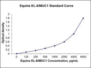
| Detection Range | 125-8000 pg/mL |
| Specificity | Equine KL-6 (MUC1) |
| Cross-Reactivity | < 0.5% cross-reactivity observed with available related molecules, < 50% cross-species reactivity observed with species tested. |
| Interference | No significant interference observed with available related molecules |
| Storage/Stability | 4 ºC for up to 6 months |
| Usage | For Laboratory Research Use Only. Not for diagnostic or therapeutic use. |
| Additional Notes | The kit allows for use in multiple experiments. |
Standard Curve
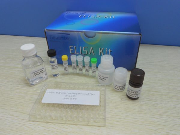
Kit Components
1. Pre-coated 96-well Microplate
2. Biotinylated Detection Antibody
3. Streptavidin-HRP Conjugate
4. Lyophilized Standards
5. TMB One-Step Substrate
6. Stop Solution
7. 20 x PBS
8. Assay Buffer
Other Materials Required but not Provided:
1. Microplate Reader capable of measuring absorption at 450 nm
2. Log-log graph paper or computer and software for ELISA data analysis
3. Precision pipettes (1-1000 µl)
4. Multi-channel pipettes (300 µl)
5. Distilled or deionized water
Protocol Outline
1. Prepare all reagents, samples and standards as instructed in the datasheet.
2. Add 100 µl of Standard or samples to each well and incubate 1 h at RT.
3. Add 100 µl of Working Detection Antibody to each well and incubate 1 h at RT.
4. Add 100 µl of Working Streptavidin-HRP to each well and incubate 20 min at RT.
5. Add 100 µl of Substrate to each well and incubate 5-30 min at RT.
6. Add 50 µl of Stop Solution to each well and read at 450 nm immediately.
Background:
Mucin 1, cell surface associated (MUC1), also called polymorphic epithelial mucin (PEM) or epithelial membrane antigen or EMA or KL-6, is a mucin encoded by the MUC1 gene.[1] MUC1 is a glycoprotein with extensive O-linked glycosylation of its extracellular domain. Mucins line the apical surface of epithelial cells in multiple organs.[2] KL-6 serves a protective function by binding to pathogens and also functions in a cell signaling capacity.[3] Overexpression of MUC1 is often associated with some cancers. Overexpression, aberrant intracellular localization, and changes in glycosylation of KL-6 have been associated with carcinomas. e.g. The CanAg tumour antigen is a novel glycoform of MUC1. In the cell nucleus, MUC1 regulates the activity of transcription factor complexes that have a role in tumor-induced changes of host immunity.[4] MUC1 cytoplasmic tail has been shown to bind to p53. This interaction is increased by genotoxic stress. MUC1 and p53 were found to be associated with the p53 response element of the p21 gene promoter. This results in activation of p21 which results in cell cycle arrest. Association of MUC1 with p53 in cancer results in inhibition of p53-mediated apoptosis and promotion of p53-mediated cell cycle arrest.[5] MUC1 cytoplasmic tail was shown to interact with Beta-catenin and this interaction was shown to be dependent on cell adhesion. A SXXXXXSSL motif was identified in MUC1 that is conserved with other beta-catenin binding partners. Studies have demonstrated that MUC1 is phosphorylated on a YEKV motif. MUC1 blocks the phosphorylation-dependent degradation of beta-catenin by GSK3B.[6] The end result is that increased expression of MUC1 in cancer increases stabilized beta-catenin. In cancer cells, increased expression of MUC1 promotes cancer cell invasion through beta-catenin, resulting in the initiation of epithelial-mesenchymal transition which promotes the formation of metastases.[7] Overexpression of MUC1 in fibroblasts increased the phosphorylation of Akt. Additionally, MUC1 was shown to increase expression of Bcl-xL.
References
- Hollingsworth MA, Swanson BJ (2004). Nature Reviews Cancer. 4 (1): 45–60.
- Moncada DM, et al. (2003). Trends Parasitol. 19 (7): 305–311.
- Lindén SK, et al. (2009). PLOS Pathog. 5 (10): e1000617.
- Li Y, Kufe D (2001). Biochem. Biophys. Res. Commun. 281 (2): 440–3.
- Raina D, Kharbanda S, Kufe D (2004). J Biol Chem. 279 (20): 20607–20612.
- Huang L, et al. (2005). Cancer Res. 65 (22): 10413–10422.
- Roy LD, et al. (2011). Oncogene. 30 (12): 1449–1459.
Be the first to review “Nori Equine KL-6 (MUC1) ELISA Kit”
You must be logged in to post a review.
Reviews
There are no reviews yet.