


Nori Equine IL-1a ELISA Kit

Price range: $508.00 through $916.00
This ELISA kit is for quantification of IL-1A in equine. This is a quick ELISA assay that reduces time to 50% compared to the conventional method, and the entire assay only takes 3 hours. This assay employs the quantitative sandwich enzyme immunoassay technique and uses biotin-streptavidin chemistry to improve the performance of the assays. An antibody specific for IL-1A has been pre-coated onto a microplate. Standards and samples are pipetted into the wells and any IL-1A present is bound by the immobilized antibody. After washing away any unbound substances, a detection antibody specific for IL-1A is added to the wells. Following wash to remove any unbound antibody reagent, a detection reagent is added. After intensive wash a substrate solution is added to the wells and color develops in proportion to the amount of IL-1A bound in the initial step. The color development is stopped, and the intensity of the color is measured.
Alternative names for IL-1a: Interleukin 1 alpha, IL1A
This product is for laboratory research use only not for diagnostic and therapeutic purposes or any other purposes.
- Description
- How Elisa Works
- Product Citations (3)
- Reviews (0)
Description
Nori Equine IL-1a ELISA Kit Summary
Alternative names for IL-1a: Interleukin 1 alpha, IL1A
Alternative names for equine: horse
| Assay Type | Solid Phase Sandwich ELISA |
| Format | 96-well Microplate or 96-Well Strip Microplate |
| Method of Detection | Colorimetric |
| Number of Targets Detected | 1 |
| Target Antigen Accession Number | Q28385 |
| Assay Length | 3 hours |
| Quantitative/Semiquantitative | Quantitative |
| Sample Type | Plasma, Serum, Cell Culture, Urine, Cell/Tissue Lysates, Synovial Fluid, BAL, |
| Recommended Sample Dilution (Plasma/Serum) | No dilution for sample <ULOQ; sufficient dilution for samples >ULOQ |
| Sensitivity | 0.5 pg/mL |
| Detection Range | 2.5-160 Pg/mL |
| Specificity | Equine IL-1a |
| Cross-Reactivity | < 0.5% cross-reactivity observed with available related molecules, < 50% cross-species reactivity observed with species tested. |
| Interference | No significant interference observed with available related molecules |
| Storage/Stability | 4 ºC for up to 6 months |
| Usage | For Laboratory Research Use Only. Not for diagnostic or therapeutic use. |
| Additional Notes | The kit allows for use in multiple experiments. |
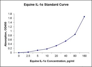
Standard Curve
Kit Components
1. Pre-coated 96-well Microplate
2. Biotinylated Detection Antibody
3. Streptavidin-HRP Conjugate
4. Lyophilized Standards
5. TMB One-Step Substrate
6. Stop Solution
7. 20 x PBS
8. Assay Buffer
Other Materials Required but not Provided:
1. Microplate Reader capable of measuring absorption at 450 nm
2. Log-log graph paper or computer and software for ELISA data analysis
3. Precision pipettes (1-1000 µl)
4. Multi-channel pipettes (300 µl)
5. Distilled or deionized water
Protocol Outline
1. Prepare all reagents, samples and standards as instructed in the datasheet.
2. Add 100 µl of Standard or samples to each well and incubate 1 h at RT.
3. Add 100 µl of Working Detection Antibody to each well and incubate 1 h at RT.
4. Add 100 µl of Working Streptavidin-HRP to each well and incubate 20 min at RT.
5. Add 100 µl of Substrate to each well and incubate 5-30 min at RT.
6. Add 50 µl of Stop Solution to each well and read at 450 nm immediately.
Background:
Interleukin-1 alpha (IL-1α) is a protein of the interleukin-1 family that in humans is encoded by the IL1A gene.[1] In general, Interleukin 1 is responsible for the production of inflammation, as well as the promotion of fever and sepsis. IL-1α inhibitors are being developed to interrupt those processes and treat diseases. IL-1α is produced mainly by activated macrophages, as well as neutrophils, epithelial cells, and endothelial cells. It possesses metabolic, physiological, haematopoietic activities, and plays one of the central roles in the regulation of the immune responses. IL1A has been shown to interact with HAX1,[2] and NDN.[3] It binds to the interleukin-1 receptor. [4] It is on the pathway that activates tumor necrosis factor-alpha. IL-1α precursor does not contain a signal peptide fragment. After processing by the removal of N-terminal amino acids by specific proteases, primarily by calpain, a calcium-activated cysteine protease.[5] Both the 31kDa precursor and 18kDa mature form of IL-1α are biologically active. It is found in substantial amounts in normal human epidermis and is distributed in a 1:1 ratio between living epidermal cells and stratum corneum.[6] The constitutive production of large amounts of IL-1α precursor by healthy epidermal keratinocytes interfere with the important role of IL-1α in immune responses, assuming skin as a barrier, which prevents the entry of pathogenic microorganisms into the body. The essential role of IL-1α in maintenance of skin barrier function, especially with increasing age,[7] is an additional explanation of IL-1α constitutive production in epidermis. IL-1α has been administered to patients during receiving autologous bone marrow transplantation.[8] The treatment with 50 ng/kg IL-1α from day zero of autologous bone marrow or stem cells transfer resulted in an earlier recovery of thrombocytopenia compared with historical controls. There is currently a head and neck phase III trial being run by Cel-Sci Corp. involving IL-1a and many other interleukins (Multikine) as an immunotherapy.
References
- March CJ, et al. (1985). Nature 315 (6021): 641–7.
- Yin H, et al. (2001). Cytokine 15 (3): 122–37.
- Hu B, et al. (2003). Natl. Acad. Sci. U.S.A. 100 (17): 10008–13.
- Bankers-Fulbright JL, Kalli KR, McKean DJ (1996). Life Sci. 59 (2): 61–83.
- Watanabe N, Kobayashi Y (1994). Cytokine 6 (6): 597–601.
- Hauser C, et al. (1986). Immunol. 136 (9): 3317–23.
- Barland CO, et al. (2004). Invest. Dermatol. 122 (2): 330–6.
- Smith JW, et al. (1993). Engl. J. Med. 328 (11): 756–61.

Product Citations
- Aziza AE et al. (2016) Effect of supplementation of broiler diets with fish oil and linseed oil on growth performance, cytokines and cecal histopathological changes in broiler chickens infected by Eimeria Tenella. Int J Agri Vet Med 4(4):12-27. Impact factor: NA. Products used and cited: Nori Chicken IL-1, IL-6 and TNF-alpha ELISA Kits.
- Mastellone V et al. (2014) Effects on oxidative status and metabolism of curcuma longa alone or associated with krill oil and ribes nigrum in healthy dogs. J Nutritional Ecol Food Res 2(4): 348-52. Impact factor: 0.6. Products used and cited: Nori Canine IL-1, IL-10 and TNFα ELISA Kits.
- Vallejo-Timaran DA et al. (2021) Pre- and post-paratam concentrations of interleukin 1α, interleukin 8 and α1-acid glycoprotein in vaginal fornix and endometrium of dairy cows with clinical cervicitis. Frontiers in Vet Sci 7,605773. Impact factor: 6.34. Products used and cited: Nori Bovine IL-1α, IL-8 and AGP ELISA Kits.
Be the first to review “Nori Equine IL-1a ELISA Kit”
You must be logged in to post a review.
Reviews
There are no reviews yet.