


Nori Bovine PDGF-AA ELISA Kit

Price range: $461.00 through $832.00
This ELISA kit is for quantification of PDGF-AA in bovine. This is a quick ELISA assay that reduces time to 50% compared to the conventional method, and the entire assay only takes 3 hours. This assay employs the quantitative sandwich enzyme immunoassay technique and uses biotin-streptavidin chemistry to improve the performance of the assays. An antibody specific for PDGF-AA has been pre-coated onto a microplate. Standards and samples are pipetted into the wells and any PDGF-AA present is bound by the immobilized antibody. After washing away any unbound substances, a detection antibody specific for PDGF-AA is added to the wells. Following wash to remove any unbound antibody reagent, a detection reagent is added. After intensive wash a substrate solution is added to the wells and color develops in proportion to the amount of PDGF-AA bound in the initial step. The color development is stopped, and the intensity of the color is measured.
Alternative names for PDGF-AA: Platelet-derived growth factor subunit A, PDGFA
This product is for Laboratory Research Use Only not for diagnostic and therapeutic purposes or any other purposes.
- Description
- How Elisa Works
- Product Citation (0)
- Reviews (0)
Description
Nori Bovine PDGF-AA ELISA Kit Summary
Alternative names for PDGF-AA: Platelet-derived growth factor subunit A, PDGFA
Alternative names for bovine: Cattle, cow, bull
| Assay Type | Solid Phase Sandwich ELISA |
| Format | 96-well Microplate or 96-Well Strip Microplate |
| Method of Detection | Colorimetric |
| Number of Targets Detected | 1 |
| Target Antigen Accession Number | Q2KJ15 |
| Assay Length | 3 hours |
| Quantitative/Semiquantitative | Quantitative |
| Sample Type | Plasma, Serum, Cell Culture, Urine, Cell/Tissue Lysates, Synovial Fluid, BAL, |
| Recommended Sample Dilution (Plasma/Serum) | No dilution for sample <ULOQ; sufficient dilution for samples >ULOQ |
| Sensitivity | 5 pg/mL |
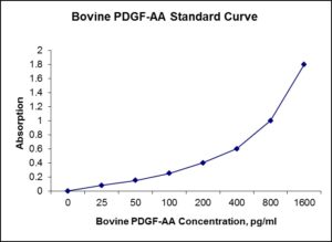
| Detection Range | 25-1600 Pg/mL |
| Specificity | Natural and recombinant bovine PDGF-AA |
| Cross-Reactivity | < 0.5% cross-reactivity observed with available related molecules, < 50% cross-species reactivity observed with species tested. |
| Interference | No significant interference observed with available related molecules |
| Storage/Stability | 4 ºC for up to 6 months |
| Usage | For Laboratory Research Use Only. Not for diagnostic or therapeutic use. |
| Additional Notes | The kit allows for use in multiple experiments. |
Standard Curve
Kit Components
1. Pre-coated 96-well Microplate
2. Biotinylated Detection Antibody
3. Streptavidin-HRP Conjugate
4. Lyophilized Standards
5. TMB One-Step Substrate
6. Stop Solution
7. 20 x PBS
8. Assay Buffer
Other Materials Required but not Provided:
1. Microplate Reader capable of measuring absorption at 450 nm
2. Log-log graph paper or computer and software for ELISA data analysis
3. Precision pipettes (1-1000 µl)
4. Multi-channel pipettes (300 µl)
5. Distilled or deionized water
Protocol Outline
1. Prepare all reagents, samples and standards as instructed in the datasheet.
2. Add 100 µl of Standard or samples to each well and incubate 1 h at RT.
3. Add 100 µl of Working Detection Antibody to each well and incubate 1 h at RT.
4. Add 100 µl of Working Streptavidin-HRP to each well and incubate 20 min at RT.
5. Add 100 µl of Substrate to each well and incubate 5-30 min at RT.
6. Add 50 µl of Stop Solution to each well and read at 450 nm immediately.
Background:
Platelet-derived growth factor subunit A is a protein that in bovines is encoded by the PDGFA gene (1,2). The protein is a member of the platelet-derived growth factor family. The four members of this family are mitogenic factors for cells of mesenchymal origin and are characterized by a motif of eight cysteines. This gene product can exist either as a homodimer or as a heterodimer with the platelet-derived growth factor beta polypeptide, where the dimers are connected by disulfide bonds. Studies using knockout mice have shown cellular defects in oligodendrocytes, alveolar smooth muscle cells, and Leydig cells in the testis; knockout mice die either as embryos or shortly after birth. Two splice variants have been identified for this gene. Though it is synthesized, stored and released by platelets upon activation, it is produced by a plethora of cells including smooth muscle cells, activated macrophages, and endothelial cells. PDGFs are mitogenic during early developmental stages, driving the proliferation of undifferentiated mesenchyme and some progenitor populations. During later maturation stages, PDGF signalling has been implicated in tissue remodelling and cellular differentiation, and in inductive events involved in patterning and morphogenesis. In addition to driving mesenchymal proliferation, PDGFs have been shown to direct the migration, differentiation and function of a variety of specialised mesenchymal and migratory cell types, both during development and in the adult animal (3).
Reference
- Stenman G, Rorsman F, Huebner K, Betsholtz C (Sep 1992). “The bovine platelet-derived growth factor alpha chain (PDGFA) gene maps to chromosome 7p22”. Cytogenet Cell Genet 60 (3–4): 206–7.
- Matsui T, Heidaran M, Miki T, Popescu N, La Rochelle W, Kraus M, Pierce J, Aaronson S (Mar 1989). “Isolation of a novel receptor cDNA establishes the existence of two PDGF receptor genes”. Science 243 (4892): 800–4.
- Hoch RV, Soriano P (2003). “Roles of PDGF in animal development”. Development 130 (20): 4769–4784.

Be the first to review “Nori Bovine PDGF-AA ELISA Kit”
You must be logged in to post a review.

Reviews
There are no reviews yet.