


Nori Bovine HGF ELISA Kit

Price range: $508.00 through $916.00
This ELISA kit is for quantification of HGF in bovine. This is a quick ELISA assay that reduces time to 50% compared to the conventional method, and the entire assay only takes 3 hours. This assay employs the quantitative sandwich enzyme immunoassay technique and uses biotin-streptavidin chemistry to improve the performance of the assays. An antibody specific for HGF has been pre-coated onto a microplate. Standards and samples are pipetted into the wells and any HGF present is bound by the immobilized antibody. After washing away any unbound substances, a detection antibody specific for HGF is added to the wells. Following wash to remove any unbound antibody reagent, a detection reagent is added. After intensive wash a substrate solution is added to the wells and color develops in proportion to the amount of HGF bound in the initial step. The color development is stopped, and the intensity of the color is measured.
Alternative names for HGF: Hepatocyte growth factor/scatter factor (HGF/SF)
This product is for Laboratory Research Use Only not for diagnostic and therapeutic purposes or any other purposes.
- Description
- How Elisa Works
- Product Citation (1)
- Reviews (0)
Description
Nori Bovine HGF ELISA Kit Summary
Alternative names for HGF: Hepatocyte growth factor/scatter factor (HGF/SF)
Alternative names for bovine: cow, cattle, bull
| Assay Type | Solid Phase Sandwich ELISA |
| Format | 96-well Microplate or 96-Well Strip Microplate |
| Method of Detection | Colorimetric |
| Number of Targets Detected | 1 |
| Accession # | Q76BS1 |
| Assay Length | 3 hours |
| Quantitative/Semiquantitative | Quantitative |
| Sample Type | Plasma, Serum, Cell Culture, Urine, Cell/Tissue Lysates, Synovial Fluid, BAL, |
| Recommended Sample Dilution (Plasma/Serum) | No dilution for sample <ULOQ; sufficient dilution for samples >ULOQ |
| Sensitivity | 18 pg/mL |
| Detection Range | 94-6000 pg/mL |
| Specificity | Bovine HGF |
| Cross-Reactivity | < 0.5% cross-reactivity observed with available related molecules, < 50% cross-species reactivity observed with species tested. |
| Interference | No significant interference observed with available related molecules |
| Storage/Stability | 4 ºC for up to 6 months |
| Usage | For Laboratory Research Use Only. Not for diagnostic or therapeutic use. |
| Additional Notes | The kit allows for use in multiple experiments. |
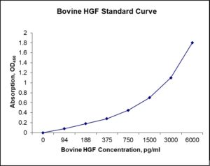
Standard Curve
Kit Components
1. Pre-coated 96-well Microplate
2. Biotinylated Detection Antibody
3. Streptavidin-HRP Conjugate
4. Lyophilized Standards
5. TMB One-Step Substrate
6. Stop Solution
7. 20 x PBS
8. Assay Buffer
Other Materials Required but not Provided:
1. Microplate Reader capable of measuring absorption at 450 nm
2. Log-log graph paper or computer and software for ELISA data analysis
3. Precision pipettes (1-1000 µl)
4. Multi-channel pipettes (300 µl)
5. Distilled or deionized water
Protocol Outline
1. Prepare all reagents, samples and standards as instructed in the datasheet.
2. Add 100 µl of Standard or samples to each well and incubate 1 h at RT.
3. Add 100 µl of Working Detection Antibody to each well and incubate 1 h at RT.
4. Add 100 µl of Working Streptavidin-HRP to each well and incubate 20 min at RT.
5. Add 100 µl of Substrate to each well and incubate 5-30 min at RT.
6. Add 50 µl of Stop Solution to each well and read at 450 nm immediately.
Background:
Hepatocyte growth factor/scatter factor (HGF/SF) is a paracrine cellular growth, motility and morphogenic factor. It is secreted by mesenchymal cells and targets and acts primarily upon epithelial cells and endothelial cells, but also acts on haemopoietic progenitor cells. It has been shown to have a major role in embryonic organ development, in adult organ regeneration and in wound healing. HGF regulates cell growth, cell motility and morphogenesis by activating a tyrosine kinase signaling cascade after binding to the proto-oncogenic c-Met receptor (1). HGF is secreted by mesenchymal cell and acts as a multifunctional cytokine on cells of mainly epithelial origin. Its ability to stimulate mitogenesis, cell motility, and matrix invasion gives it a central role in angiogenesis, tumorogenesis, and tissue regeneration. It is secreted as a single inactive polypeptide and is cleaved by serine protease into a 69-kDa alpha-chain and 34-kDa beta-chain. A disulfide bond between the alpha and beta chains produces the active, heterodimeric molecule. The protein belongs to the plasminogen subfamily of S1 peptidases but has no detectable protease activity (2). Human HGF plasmid DNA therapy of cardiomycytes is being examined as a potential treatment for coronary artery disease as well as treatment for the damage that occurs to the heart after myocardial infarction (3, 4). HGF may further play a role as an indicator for prognosis of chronicity for Chikungunya virus induced arthralgia. High HGF levels correlate with high rate of recovery (5).
Reference
- DP Bottaro et al. (1991) Science 251 (4995) : 802.
- T Nakamura et al. (1989) Nature 342 : 440.
- ZJ Yang et al. (2008) Molecular Biology Reports 36(6) : 1323.
- W Hahn et al. (2011) The Journal of Gene Medicine 13 (10) : 549.
- A Chow et al. (2011) J Infect. Dis. 203(2) : 149.

Product Citations
- Jones AK et al. (2017) Gestational restricted- and over-feeding promote maternal and offspring inflammatory response that are distinct and dependent on diet on sheep. Biology of Reproduction doi:10.1093/biolre/iox174. Impact factor: 3.714. Products used and cited: Nori Sheep BMP2, HGF, TGFb1 ELISA Kits.
Be the first to review “Nori Bovine HGF ELISA Kit”
You must be logged in to post a review.
Reviews
There are no reviews yet.