Nori Hamster IgM ELISA Kit
$461.00 – $832.00
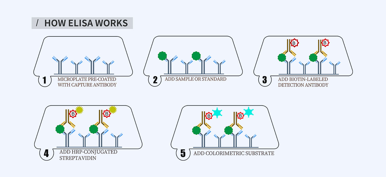
This ELISA kit is for quantification of IgM in hamster. This is a quick ELISA assay that reduces time to 50% compared to the conventional method, and the entire assay only takes 3 hours. This assay employs the quantitative sandwich enzyme immunoassay technique and uses biotin-streptavidin chemistry to improve the performance of the assays. An antibody specific for IgM has been pre-coated onto a microplate. Standards and samples are pipetted into the wells and any IgM present is bound by the immobilized antibody. After washing away any unbound substances, a detection antibody specific for IgM is added to the wells. Following wash to remove any unbound antibody reagent, a detection reagent is added. After intensive wash a substrate solution is added to the wells and color develops in proportion to the amount of IgM bound in the initial step. The color development is stopped, and the intensity of the color is measured.
Alternative names for IgM: Immunoglobulin M
This product is for Laboratory Research Use Only not for diagnostic and therapeutic purposes or any other purposes.
- Description
- How Elisa Works
- Product Citation (0)
- Reviews (0)
Description
Nori Hamster IgM ELISA Kit Summary
Alternative names for IgM: Immunoglobulin M
Alternative names for hamster: Golden hamster, syrian hamster
| Assay Type | Solid Phase Sandwich ELISA |
| Format | 96-well Microplate or 96-Well Strip Microplate |
| Method of Detection | Colorimetric |
| Number of Targets Detected | 1 |
| Target Antigen Accession Number | na |
| Assay Length | 3 hours |
| Quantitative/Semiquantitative | Quantitative |
| Sample Type | Plasma, Serum, Cell Culture, Urine, Cell/Tissue Lysates, Synovial Fluid, BAL, |
| Recommended Sample Dilution (Plasma/Serum) | No dilution for sample <ULOQ; sufficient dilution for samples >ULOQ |
| Sensitivity | 600 pg/mL |
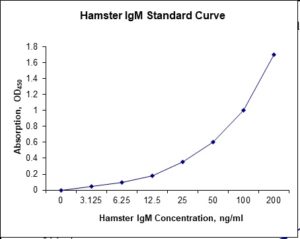
| Detection Range | 3.125-200 ng/mL |
| Specificity | Natural and recombinant hamster IgM |
| Cross-Reactivity | < 0.5% cross-reactivity observed with available related molecules, < 50% cross-species reactivity observed with species tested. |
| Interference | No significant interference observed with available related molecules |
| Storage/Stability | 4 ºC for up to 6 months |
| Usage | For Laboratory Research Use Only. Not for diagnostic or therapeutic use. |
| Additional Notes | The kit allows for use in multiple experiments. |
Standard Curve
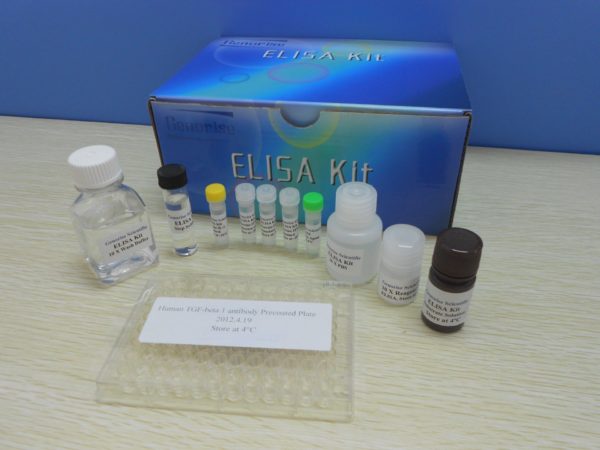
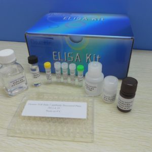
Kit Components
1. Pre-coated 96-well Microplate
2. Biotinylated Detection Antibody
3. Streptavidin-HRP Conjugate
4. Lyophilized Standards
5. TMB One-Step Substrate
6. Stop Solution
7. 20 x PBS
8. Assay Buffer
Other Materials Required but not Provided:
1. Microplate Reader capable of measuring absorption at 450 nm
2. Log-log graph paper or computer and software for ELISA data analysis
3. Precision pipettes (1-1000 µl)
4. Multi-channel pipettes (300 µl)
5. Distilled or deionized water
Protocol Outline
1. Prepare all reagents, samples and standards as instructed in the datasheet.
2. Add 100 µl of Standard or samples to each well and incubate 1 h at RT.
3. Add 100 µl of Working Detection Antibody to each well and incubate 1 h at RT.
4. Add 100 µl of Working Streptavidin-HRP to each well and incubate 20 min at RT.
5. Add 100 µl of Substrate to each well and incubate 5-30 min at RT.
6. Add 50 µl of Stop Solution to each well and read at 450 nm immediately.
Background:
Immunoglobulin M (IgM) is a basic antibody that is produced by B cells. IgM is by far the physically largest antibody in the human circulatory system. It is the first antibody to appear in response to initial exposure to an antigen. The spleen is the major site of specific IgM production. IgM forms polymers where multiple immunoglobulins are covalently linked together with disulfide bonds, mostly as a pentamer but also as a hexamer. The J chain is found in pentameric IgM but not in the hexameric form.[1] IgM has a molecular mass of approximately 970 kDa (in its pentamer form). Because each monomer has two antigen binding sites, a pentameric IgM has 10 binding sites. However, IgM cannot bind 10 antigens at the same time because the large size of most antigens hinders binding to nearby sites. Because IgM is a large molecule, it cannot diffuse well, and is found in the interstitium only in very low quantities. IgM is primarily found in serum; however, because of the J chain, it is also important as a secretory immunoglobulin. Due to its polymeric nature, IgM possesses high avidity, and is particularly effective at complement activation. By itself, IgM is an ineffective opsonin; however it contributes greatly to opsonization by activating complement and causing C3b to bind to the antigen.[2] IgM antibodies appear early in the course of an infection and usually reappear, to a lesser extent, after further exposure. IgM antibodies do not pass across the human placenta (only isotype IgG). These two biological properties of IgM make it useful in the diagnosis of infectious diseases. Demonstrating IgM antibodies in a patient’s serum indicates recent infection, or in a neonate’s serum indicates intrauterine infection (e.g. congenital rubella). The development of anti-donor IgM after organ transplantation is not associated with graft rejection but it may have a protective effect.[3]
References
- Erik J. et al, J. Immunol., Jun 1998; 160: 5979 – 5989.
- Wellek, B, et al. (1976). Agents and Actions 6 (1–3): 260–262.
- Charpak, Y, et al. (2004). Liver Transpl 10 (2): 315–319.
Be the first to review “Nori Hamster IgM ELISA Kit”
You must be logged in to post a review.




Reviews
There are no reviews yet.