





















Nori Rat TERF2 ELISA Kit
Price range: $508.00 through $916.00
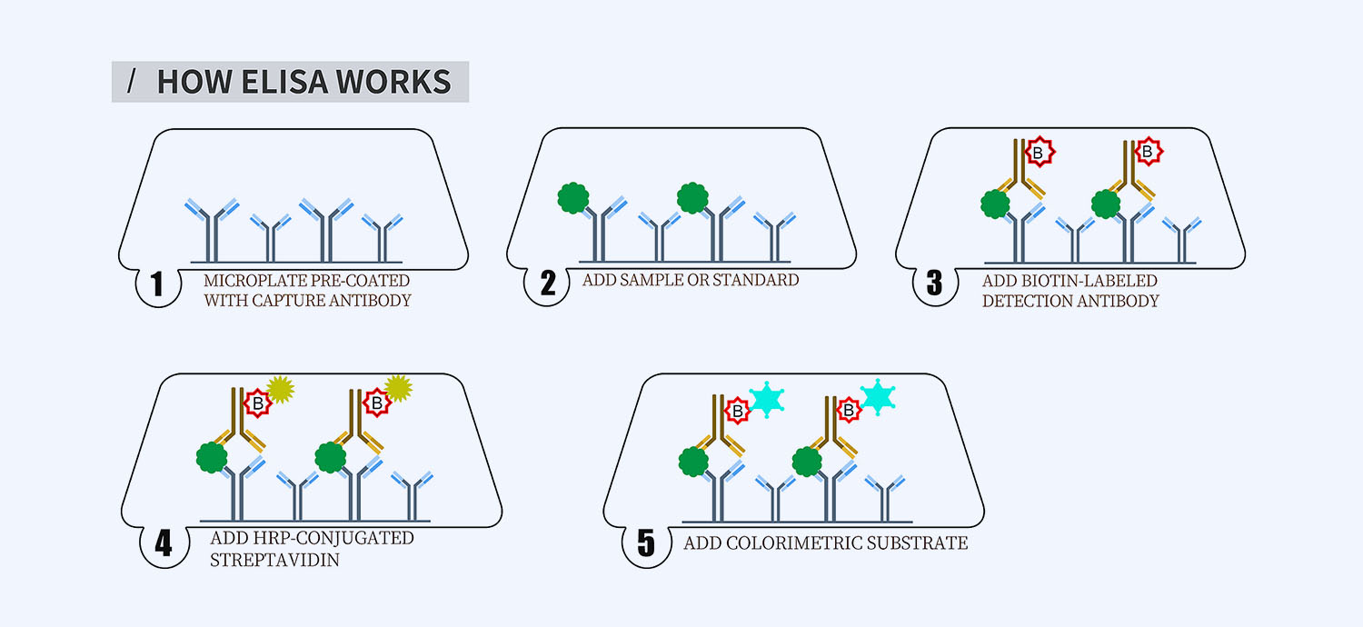
This ELISA kit is for quantification of TERF2 in rat. This is a quick ELISA assay that reduces time to 50% compared to the conventional method, and the entire assay only takes 3 hours. This assay employs the quantitative sandwich enzyme immunoassay technique and uses biotin-streptavidin chemistry to improve the performance of the assays. An antibody specific for TERF2 has been pre-coated onto a microplate. Standards and samples are pipetted into the wells and any TERF2 present is bound by the immobilized antibody. After washing away any unbound substances, a detection antibody specific for TERF2 is added to the wells. Following wash to remove any unbound antibody reagent, a detection reagent is added. After intensive wash a substrate solution is added to the wells and color develops in proportion to the amount of TERF2 bound in the initial step. The color development is stopped, and the intensity of the color is measured.
Alternative names for TERF2: Telomeric repeat-binding factor 2 , TRF2, TRBF2
This product is for Laboratory Research Use Only not for diagnostic and therapeutic purposes or any other purposes.
- Description
- How Elisa Works
- Documents
- Product Citation (0)
- Reviews (0)
Description
Nori Rat TERF2 ELISA Kit Summary
Alternative names for TERF2: Telomeric repeat-binding factor 2 , TRF2, TRBF2
| Assay Type | Solid Phase Sandwich ELISA |
| Format | 96-well Microplate or 96-Well Strip Microplate |
| Method of Detection | Colorimetric |
| Number of Targets Detected | 1 |
| Target Antigen Accession Number |
D3ZJF7 |
| Assay Length | 3 hours |
| Quantitative/Semiquantitative | Quantitative |
| Sample Type | Plasma, Serum, Cell Culture, Urine, Cell/Tissue Lysates, Synovial Fluid, BAL, |
| Recommended Sample Dilution (Plasma/Serum) | No dilution for sample <ULOQ; sufficient dilution for samples >ULOQ |
| Sensitivity | 30 pg/mL |
| Detection Range | 0.156-10 ng/mL |
| Specificity | Rat TERF2 |
| Cross-Reactivity | < 0.5% cross-reactivity observed with available related molecules, < 50% cross-species reactivity observed with species tested. |
| Interference | No significant interference observed with available related molecules |
| Storage/Stability | 4 ºC for up to 6 months |
| Usage | For Laboratory Research Use Only. Not for diagnostic or therapeutic use. |
| Additional Notes | The kit allows for use in multiple experiments. |
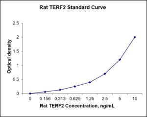
Standard Curve
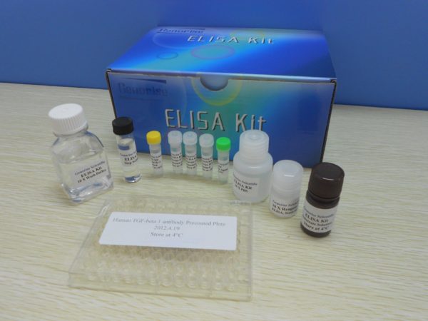
Kit Components
1. Pre-coated 96-well Microplate
2. Biotinylated Detection Antibody
3. Streptavidin-HRP Conjugate
4. Lyophilized Standards
5. TMB One-Step Substrate
6. Stop Solution
7. 20 x PBS
8. Assay Buffer
Other Materials Required but not Provided:
1. Microplate Reader capable of measuring absorption at 450 nm
2. Log-log graph paper or computer and software for ELISA data analysis
3. Precision pipettes (1-1000 µl)
4. Multi-channel pipettes (300 µl)
5. Distilled or deionized water
Protocol Outline
1. Prepare all reagents, samples and standards as instructed in the datasheet.
2. Add 100 µl of Standard or samples to each well and incubate 1 h at RT.
3. Add 100 µl of Working Detection Antibody to each well and incubate 1 h at RT.
4. Add 100 µl of Working Streptavidin-HRP to each well and incubate 20 min at RT.
5. Add 100 µl of Substrate to each well and incubate 5-30 min at RT.
6. Add 50 µl of Stop Solution to each well and read at 450 nm immediately.
Background:
Telomeric repeat-binding factor 2 is a protein that is present at telomeres throughout the cell cycle and plays a key role in the protective activity of telomeres. It is also known as TERF2, TRF2, and TRBF2, and is encoded by the TERF2 gene. It is a component of the shelterin nucleoprotein complex and a second negative regulator of telomere length, playing a key role in the protective activity of telomeres. TERF2 differs from TERF1 in that its N terminus is basic rather than acidic. TERF2 promotes t-loop formation by preferentially binding to a telomeric double-stranded DNA duplex containing a 3’ TTAGGG single-stranded overhang. Interaction of TERF2 with TERF2IP is shown to promote higher t-loop formation in vitro.[1] Studies have demonstrated that deletion of TERF2 prevents t-loop formation, leading to excessive loss of telomeric DNA and early cell death.[2] TERF2 plays a central role in preventing ATM kinase DNA damage response. It binds telomeric dsDNA and prevents telomeres from activating ATM kinase. Removal of TERF2 induces ATM-dependent apoptosis by localizing the active, phosphorylated form of ATM to unprotected chromosome ends.[3] TERF2 is also known to recruit certain client proteins, also known as accessory factors. These client proteins are often recruited to TERF2 for a specific function at a specific time, often temporarily. TERF2 as part of the shelterin complex, has been known to block the ATM signaling pathways and prevent chromosome end fusion. In cancer cells, TERF2 phosphorylation by ERK1/2 is a controlling factor in the major pro-oncogenic signaling pathways (RAS/RAF/MEK/ERK) that affect telomeric stability.[4] Additionally, when TERF2 was non-phosphorylated in melanoma cells, there was a cell induced DNA damage response, arresting growth and causing tumor reversion.[4] Studies have found that in tumor cells, TERF2 levels are observed to be high, and this raised level of TERF2 contributes to oncogenesis in a variety of ways.[23][24][25] This high level of TERF2 decreases the ability to recruit and activate natural killer cells in human tumor cells.[5] One study used a dominant negative form of TERF2ΔBΔC, to inhibit TERF2, and found that it could induce a reversion malignant phenotype in human melanoma cells.[6] Therefore, over-expression of TERF2ΔBΔC, and therefore blocking of TERF2, induced apoptosis and reduced tumourigenicity in certain cell lines.[24] Additionally, upregulation of TERF2 may be the cause of the establishment and maintenance of short telomeres.[7] These short telomeres increase chromosomal instability, and increase the chances of certain cancers progressing in the body, such as with leukemia.[7] In gastric mucosa tissues, the expression of TERF2 proteins was significantly higher than normal, and this over-expression of TERF2, along with over-expression of TERF1, TIN2, TERT, and BRCA1 protein transposition, may cause a reduction in telomere length, further contributing to multistage carcinogenesis of gastric cancer.[8]
References
- Arat NÖ, Griffith JD (2012). The Journal of Biological Chemistry. 287 (50): 41583–94.
- Stansel RM, de Lange T, Griffith JD (2001). The EMBO Journal. 20 (19): 5532–40.
- Karlseder J, et al. (2004). PLoS Biology. 2 (8): E240. doi:1371/journal.pbio.0020240.
- Picco V, et al. (2016). Oncotarget. 7 (29): 46615–46627.
- Biroccio A, et al. (2013). Nature Cell Biology. 15 (7): 818–28. doi:1038/ncb2774. PMID23792691.
- Biroccio A, et al. (2006). European Journal of Cancer. 42 (12): 1881–8.
- Bellon M, et al. (2006). International Journal of Cancer. 119 (9): 2090–7.
- Hu H, et al. (2010). Journal of Cancer Research and Clinical Oncology. 136 (9): 1407–14.
Be the first to review “Nori Rat TERF2 ELISA Kit”
You must be logged in to post a review.
Reviews
There are no reviews yet.