


Nori Canine CA-125 ELISA Kit

Price range: $508.00 through $916.00
This ELISA kit is for quantification of CA-125 in canine. This is a quick ELISA assay that reduces time to 50% compared to the conventional method, and the entire assay only takes 3 hours. This assay employs the quantitative sandwich enzyme immunoassay technique and uses biotin-streptavidin chemistry to improve the performance of the assays. An antibody specific for CA-125 has been pre-coated onto a microplate. Standards and samples are pipetted into the wells and any CA-125 present is bound by the immobilized antibody. After washing away any unbound substances, a detection antibody specific for CA-125 is added to the wells. Following wash to remove any unbound antibody reagent, a detection reagent is added. After intensive wash a substrate solution is added to the wells and color develops in proportion to the amount of CA-125 bound in the initial step. The color development is stopped, and the intensity of the color is measured.
Alternative names for CA-125: carcinoma antigen 125, carbohydrate antigen125, mucin 16, MUC16, CA125
This product is for laboratory research use only not for diagnostic and therapeutic purposes or any other purposes.
- Description
- How Elisa Works
- Documents
- Product Citations
- Reviews (0)
Description
Nori Canine CA-125 II ELISA Kit Summary
Alternative names for CA-125: carcinoma antigen 125, carbohydrate antigen125, mucin 16, MUC16, CA125
Alternative names for canine: Dog
| Assay Type | Solid Phase Sandwich ELISA |
| Format | 96-well Microplate or 96-Well Strip Microplate |
| Method of Detection | Colorimetric |
| Number of Targets Detected | 1 |
| Target Antigen Accession Number | A0A140LJ72 |
| Assay Length | 3 hours |
| Quantitative/Semiquantitative | Quantitative |
| Sample Type | Plasma, Serum, Cell Culture, Urine, Cell/Tissue Lysates, Synovial Fluid, BAL, |
| Recommended Sample Dilution (Plasma/Serum) | No dilution for sample <ULOQ; sufficient dilution for samples >ULOQ |
| Sensitivity | 1.2 U/mL |
| Detection Range | 6.25-400 U/mL |
| Specificity | Canine CA-125 |
| Cross-Reactivity | < 0.5% cross-reactivity observed with available related molecules, < 50% cross-species reactivity observed with species tested. |
| Interference | No significant interference observed with available related molecules |
| Storage/Stability | 4 ºC for up to 6 months |
| Usage | For Laboratory Research Use Only. Not for diagnostic or therapeutic use. |
| Additional Notes | The kit allows for use in multiple experiments. |
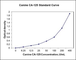
Standard Curve
Kit Components
1. Pre-coated 96-well Microplate
2. Biotinylated Detection Antibody
3. Streptavidin-HRP Conjugate
4. Lyophilized Standards
5. TMB One-Step Substrate
6. Stop Solution
7. 20 x PBS
8. Assay Buffer
Other Materials Required but not Provided:
1. Microplate Reader capable of measuring absorption at 450 nm
2. Log-log graph paper or computer and software for ELISA data analysis
3. Precision pipettes (1-1000 µl)
4. Multi-channel pipettes (300 µl)
5. Distilled or deionized water
Protocol Outline
1. Prepare all reagents, samples and standards as instructed in the datasheet.
2. Add 100 µl of Standard or samples to each well and incubate 1 h at RT.
3. Add 100 µl of Working Detection Antibody to each well and incubate 1 h at RT.
4. Add 100 µl of Working Streptavidin-HRP to each well and incubate 20 min at RT.
5. Add 100 µl of Substrate to each well and incubate 5-30 min at RT.
6. Add 50 µl of Stop Solution to each well and read at 450 nm immediately.
Background:
CA-125 (carcinoma antigen 125, or carbohydrate antigen 125) also known as mucin 16 (MUC16) is a protein that is encoded by the MUC16 gene.[1] MUC16 is a member of the mucin family and is a membrane associated muci. and a unique property of MUC16 is its large size (22,000 amino acids) and is more than twice than MUC1 and MUC4, making it the largest membrane-associated mucin. The N-terminal and tandem repeat domains are both entirely extracellular and highly O-glycosylated. The C-terminal domain contains multiple extracellular SEA modules, a transmembrane domain, and a cytoplasmic tail. The extracellular region of MUC16 can be released from the cell surface by undergoing proteolytic cleavage. MUC16 is thought to be cleaved at a site in the SEA modules. MUC16 is a component of the ocular surface, the respiratory tract and the female reproductive tract epithelia. Since MUC16 is highly glycosylated it creates a hydrophilic environment that acts as a lubricating barrier against foreign particles and infectious agents on the apical membrane of epithelial cells. Also, the cytoplasmic tail of MUC16 has been shown to interact with cytoskeleton by binding members of the ERM protein family.[2] The expression of mucin 16 has been shown to be altered in dry eye, cystic fibrosis, and several types of cancers. CA-125 may play a role in advancing tumorigenesis and tumor proliferation by several different mechanisms. MUC16 is thought to participate in cell-to-cell interactions that enable the metastasis of tumor cells. MUC16 binds selectively to mesothelin at the N-terminus of cell surface mesothelin.[3] MUC16 and mesothelin interactions are thought to provide the first step in tumor cell invasion of the peritoneum.[4] An immunoadhesin (HN125) has the ability to disrupt the heterotypic cancer cell adhesion mediated by the MUC16-mesothelin interaction.[24] MUC16 and mesothelial interactions may aid in the gathering of other tumor cells to the location of a metastasis, thus increasing the size of the metastasis.[4] Evidence suggests that expression of the cytoplasmic tail of MUC16 enables tumor cells to grow, promotes cell motility and may facilitate invasion. MUC16 may reduce the sensitivity of cancer cells to drug therapy. CA-125 may serve as a tumor marker since it is elevated in the blood of some patients with specific types of cancers, most notably ovarian cancer, or other conditions that are benign. Testing of CA-125 blood levels has been proposed as useful in treating ovarian cancer. While the test can give useful information for women already known to have ovarian cancer, CA-125 testing has not been found useful as a screening method because of the uncertain correlation between CA-125 levels and cancer. In addition to ovarian cancer, CA-125 can be elevated in other types of cancers and in pregnant women. Because of the wide variety of conditions that can increase serum levels, CA-125 is not used to detect cancer, but it is often used to monitor responses to chemotherapy, relapse, and disease progression in ovarian cancer patients.
References
- Yin BW, et al. (2002). International Journal of Cancer. 98(5): 737–40.
- Blalock TD, et al. (2007). Investigative Ophthalmology & Visual Science. 48(10): 4509–18.
- Rump A, et al. (2004). The Journal of Biological Chemistry. 279(10): 9190–8.
- Gubbels JA, et al. (2006). Molecular Cancer. 5(1): 50.

Product Citations
Be the first to review “Nori Canine CA-125 ELISA Kit”
You must be logged in to post a review.
Reviews
There are no reviews yet.