


Nori Rabbit Total Cathepsin S ELISA Kit

Price range: $508.00 through $916.00
This ELISA kit is for quantification of total CTSS in rabbit. This is a quick ELISA assay that reduces time to 50% compared to the conventional method, and the entire assay only takes 3 hours. This assay employs the quantitative sandwich enzyme immunoassay technique and uses biotin-streptavidin chemistry to improve the performance of the assays. An antibody specific for CTSS has been pre-coated onto a microplate. Standards and samples are pipetted into the wells and any CTSS present is bound by the immobilized antibody. After washing away any unbound substances, a detection antibody specific for CTSS is added to the wells. Following wash to remove any unbound antibody reagent, a detection reagent is added. After intensive wash a substrate solution is added to the wells and color develops in proportion to the amount of CTSS bound in the initial step. The color development is stopped, and the intensity of the color is measured.
Alternative names for cathepsin S: CTSS
This product is for laboratory research use only not for diagnostic and therapeutic purposes or any other purposes.
- Description
- How Elisa Works
- Product Citation (0)
- Reviews (0)
Description
Nori Rabbit Total Cathepin S ELISA Kit Summary
Alternative names for cathepsin S: CTSS,
| Assay Type | Solid Phase Sandwich ELISA |
| Format | 96-well Microplate or 96-Well Strip Microplate |
| Method of Detection | Colorimetric |
| Number of Targets Detected | 1 |
| Target Antigen Accession Number | G1T0K7 |
| Assay Length | 3 hours |
| Quantitative/Semiquantitative | Quantitative |
| Sample Type | Plasma, Serum, Cell Culture, Urine, Cell/Tissue Lysates, Synovial Fluid, BAL, |
| Recommended Sample Dilution (Plasma/Serum) | No dilution for sample <ULOQ; sufficient dilution for samples >ULOQ |
| Sensitivity | 30 pg/mL |
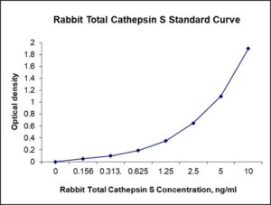
| Detection Range | 0.156-10 ng/mL |
| Specificity | Rabbit cathepsin S |
| Cross-Reactivity | < 0.5% cross-reactivity observed with available related molecules, < 50% cross-species reactivity observed with species tested. |
| Interference | No significant interference observed with available related molecules |
| Storage/Stability | 4 ºC for up to 6 months |
| Usage | For Laboratory Research Use Only. Not for diagnostic or therapeutic use. |
| Additional Notes | The kit allows for use in multiple experiments. |
Standard Curve
Kit Components
1. Pre-coated 96-well Microplate
2. Biotinylated Detection Antibody
3. Streptavidin-HRP Conjugate
4. Lyophilized Standards
5. TMB One-Step Substrate
6. Stop Solution
7. 20 x PBS
8. Assay Buffer
Other Materials Required but not Provided:
1. Microplate Reader capable of measuring absorption at 450 nm
2. Log-log graph paper or computer and software for ELISA data analysis
3. Precision pipettes (1-1000 µl)
4. Multi-channel pipettes (300 µl)
5. Distilled or deionized water
Protocol Outline
1. Prepare all reagents, samples and standards as instructed in the datasheet.
2. Add 100 µl of Standard or samples to each well and incubate 1 h at RT.
3. Add 100 µl of Working Detection Antibody to each well and incubate 1 h at RT.
4. Add 100 µl of Working Streptavidin-HRP to each well and incubate 20 min at RT.
5. Add 100 µl of Substrate to each well and incubate 5-30 min at RT.
6. Add 50 µl of Stop Solution to each well and read at 450 nm immediately.
Background:
Cathepsin S is a protein that is encoded by the CTSS gene.[1] Cathepsin S is a member of the peptidase C1 family and is a lysosomal cysteine protease that may participate in the degradation of antigenic proteins to peptides for presentation to the MHC class II. Cathepsin S can function as an elastase over a broad pH range in alveolar macrophages. Cathepsin S is a lysosomal enzyme that belongs to the papain family of cysteine proteases. While a role in antigen presentation has long been recognized, it is now understood that cathepsin S has a role in itch and pain, or nociception. The mechanism by which cathepsin S leads to itch and pain is consistent with the capacity of this cysteine protease to activate protease-activated receptors 2 and 4.[2][3] Cathepsin S is expressed by antigen presenting cells including macrophages, B-lymphocytes, dendritic cells, microglia and some epithelial cells. Its expression is markedly increased in human keratinocytes following stimulation with interferon-gamma and its expression is elevated in psoriatic keratinocytes due to stimulation by proinflammatory factors. In contrast, cortical thymic epithelial cells do not express cathepsin S. Immune cells, including macrophages and microglia, secrete cathepsin S in response to inflammatory mediators including lipopolysaccharides, proinflammatory cytokines and neutrophils. The expression of cathepsin S can be triggered by proinflammatory factors secreted by tumor cells. Cathepsin S specially cleaves and activates the psoriasis-associated proinflammatory cytokine IL-36γ[7] In vitro, cathepsin S retains some enzyme activity in the presence of 3M urea. Cathepsin S is produced as a zymogen and is activated by processing. The activity of cathepsin S is tightly regulated by its endogenous inhibitor, cystatin C, which also has a role in antigen presentation. Cystatin A and B have a lower activity compared to cystatin C. Cathepsin S participates in the degradation of the invariant or Ii chain that prevents loading the antigen into the complex. Cathepsin S has been shown to be a significant prognostic factor for patients with type IV astrocytomas.
References
- Shi GP, et al. (1994). The Journal of Biological Chemistry. 269 (15): 11530–6.
- Reddy VB, et al. (2015). Nature Communications. 6: 7864. doi:1038/ncomms8864.
- Elmariah SB, et al. (2014). PLOS ONE. 9 (6): e99702. doi:1371/journal.pone.0099702.
- Ainscough JS, et al. (2017). Proc Natl Acad Sci USA. 114 (13): E2748–E2757. doi:1073/pnas.1620954114.

Be the first to review “Nori Rabbit Total Cathepsin S ELISA Kit”
You must be logged in to post a review.

Reviews
There are no reviews yet.