


Nori Porcine S100A11 ELISA Kit

$461.00 – $832.00
DataSheet CoA SDS
This ELISA kit is for quantification of S100A11 in pig. This is a quick ELISA assay that reduces time to 50% compared to the conventional method, and the entire assay only takes 3 hours. This assay employs the quantitative sandwich enzyme immunoassay technique and uses biotin-streptavidin chemistry to improve the performance of the assays. An antibody specific for S100A11 has been pre-coated onto a microplate. Standards and samples are pipetted into the wells and any S100A11 present is bound by the immobilized antibody. After washing away any unbound substances, a detection antibody specific for S100A11 is added to the wells. Following wash to remove any unbound antibody reagent, a detection reagent is added. After intensive wash a substrate solution is added to the wells and color develops in proportion to the amount of S100A11 bound in the initial step. The color development is stopped, and the intensity of the color is measured.
Alternative names for S100A11: S100 calcium-binding protein A11, S100-A11, S100C, calgizzarin
This product is for Laboratory Research Use Only not for diagnostic and therapeutic purposes or any other purposes.
- Description
- How Elisa Works
- Product Citation (0)
- Reviews (0)
Description
Nori Porcine S100A11 ELISA Kit Summary
Alternative names for S100A11: S100 calcium-binding protein A11, S100-A11, calgizzarin or S100C
Alternative name for porcine: Pig
| Assay Type | Solid Phase Sandwich ELISA |
| Format | 96-well Microplate or 96-Well Strip Microplate |
| Method of Detection | Colorimetric |
| Number of Targets Detected | 1 |
| Target Antigen Accession Number | P31950 |
| Assay Length | 3 hours |
| Quantitative/Semiquantitative | Quantitative |
| Sample Type | Plasma, Serum, Cell Culture, Urine, Cell/Tissue Lysates, Synovial Fluid, BAL, |
| Recommended Sample Dilution (Plasma/Serum) | No dilution for sample <ULOQ; sufficient dilution for samples >ULOQ |
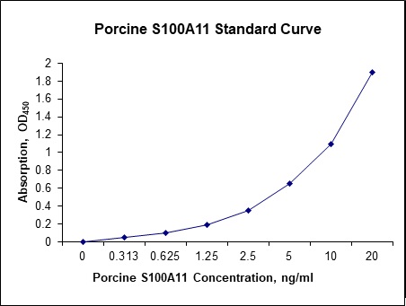
| Sensitivity | 60 pg/mL |
| Detection Range | 0.31-20 ng/mL |
| Specificity | Natural and recombinant porcine S100A11 |
| Cross-Reactivity | < 0.5% cross-reactivity observed with available related molecules, < 50% cross-species reactivity observed with species tested. |
| Interference | No significant interference observed with available related molecules |
| Storage/Stability | 4 ºC for up to 6 months |
| Usage | For Laboratory Research Use Only. Not for diagnostic or therapeutic use. |
| Additional Notes | The kit allows for use in multiple experiments. |
Standard Curve
Kit Components
1. Pre-coated 96-well Microplate
2. Biotinylated Detection Antibody
3. Streptavidin-HRP Conjugate
4. Lyophilized Standards
5. TMB One-Step Substrate
6. Stop Solution
7. 20 x PBS
8. Assay Buffer
Other Materials Required but not Provided:
1. Microplate Reader capable of measuring absorption at 450 nm
2. Log-log graph paper or computer and software for ELISA data analysis
3. Precision pipettes (1-1000 µl)
4. Multi-channel pipettes (300 µl)
5. Distilled or deionized water
Protocol Outline
1. Prepare all reagents, samples and standards as instructed in the datasheet.
2. Add 100 µl of Standard or samples to each well and incubate 1 h at RT.
3. Add 100 µl of Working Detection Antibody to each well and incubate 1 h at RT.
4. Add 100 µl of Working Streptavidin-HRP to each well and incubate 20 min at RT.
5. Add 100 µl of Substrate to each well and incubate 5-30 min at RT.
6. Add 50 µl of Stop Solution to each well and read at 450 nm immediately.
Background:
S100 calcium-binding protein A11 (S100A11) is a protein that is encoded by the S100A11 gene. S100A11 is a member of the S100 family of proteins containing 2 EF-hand calcium-binding motifs. S100A11, also known as calgizzarin or 100C, is a small acidic protein. Usually, S100A11 is in homodimeres, but it has been shown that S100A11 heterodimerizes with S100B[1] and it also interacts with Nucleolin,[2] and RAD54B.[3] The protein may function in motility, invasion, and tubulin polymerization. Chromosomal rearrangements and altered expression of S100A11 have been implicated in tumor metastasis. S100A11 is highly expressed in many tissues.[4] This protein is normally found strictly in the nucleus, but appears in the cytoplasm in cancer cells. S100A11 was localized in the cytoplasm of resting human keratinocytes in vitro.[5] S100A11 is implicated in membrane and cytoskeletar dynamics, vesicular transportation and processes of endo and exocytosis. S100A11 may interact with many cytoskeletal structures.[5] S100A11 is able to control reorganization of actin and it is important in forming protrusion by metastatic cells. S100A11 interacts with the RAGE receptor, which is also a receptor for other S100 proteins. It lacks enzymatic activity, but regulates activity of other enzymes and functions by binding to other proteins.[6] It is associated with cell cycle, growth, survival and apoptosis. It has been identified as dual growth mediator. Suppression of S100A11 by small interfering RNA caused cells to apoptosis, and overexpression of S100A11 has been found to inhibit apoptosis in tumor cells. Furthermore, the knock-down of S100A11 via siRNA reduces the sister-chromatid exchange and the viability of cells. IL-8 and TNF-alpha induce the expression and release of S100A11 in chondrocytes in culture and exogenous S100A11 causes chondrocyte hypertrophy. S100A11 could play a role in maintaining low-grade inflammation in osteoarthritis and in its progression.[7] It is associated with many different types of cancers. Its overproduction has been found, for example, in breast, pancreas or colectal carcinoma and its levels can be used as clinical marker in these diseases.[5] S100A11 enhances the recombination activity of human RAD51 in vitro. A knock-down leads to diffuse distribution of RAD54B, suggesting a potential role of S100A11 in the process of homologous recombination repair of double-strand breaks.[8]
References
- Deloulme JC, et al. (2000). The Journal of Biological Chemistry. 275(45): 35302–10.
- Sakaguchi M, et al. (2003). The Journal of Cell Biology. 163(4): 825–35.
- Murzik U, et al. (2008). Molecular Biology of the Cell. 19(7): 2926–35.
- Inada H, et al. (1999). Biochemical and Biophysical Research Communications. 263(1): 135–8.
- Sakaguchi M, Huh NH (2011). Amino Acids. 41(4): 797–807.
- Zhao XQ, et al. (2000). Biochemical and Biophysical Research Communications. 267(1): 77–9.
- Cecil DL, Terkeltaub R (2008). Journal of Immunology. 180(12): 8378–85.
- Foertsch F, et al. (2016). Cell Cycle. 15(20): 2766–79.

Be the first to review “Nori Porcine S100A11 ELISA Kit”
You must be logged in to post a review.
Reviews
There are no reviews yet.