Nori Mouse MPO ELISA Kit
$461.00 – $832.00
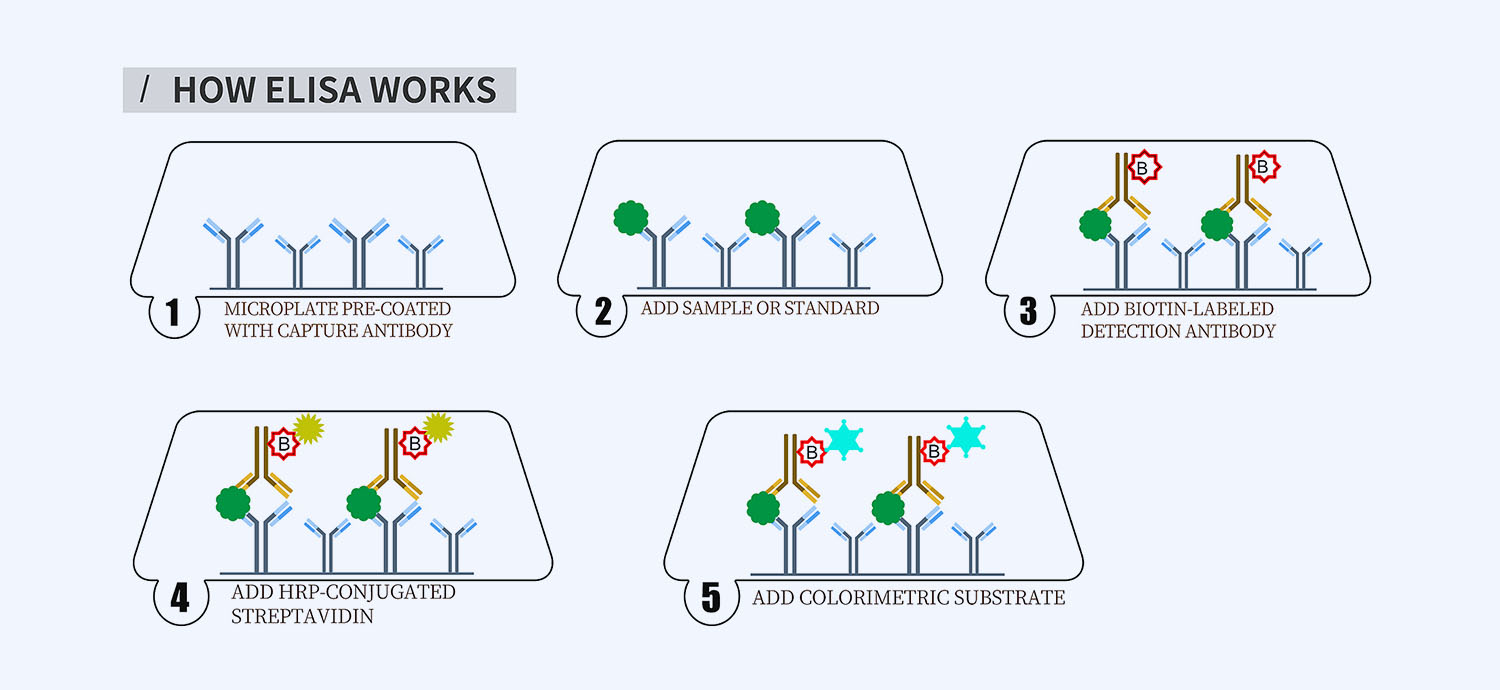
This ELISA kit is for quantification of MPO in mouse. This is a quick ELISA assay that reduces time to 50% compared to the conventional method, and the entire assay only takes 3 hours. This assay employs the quantitative sandwich enzyme immunoassay technique and uses biotin-streptavidin chemistry to improve the performance of the assays. An antibody specific for MPO has been pre-coated onto a microplate. Standards and samples are pipetted into the wells and any MPO present is bound by the immobilized antibody. After washing away any unbound substances, a detection antibody specific for MPO is added to the wells. Following wash to remove any unbound antibody reagent, a detection reagent is added. After intensive wash a substrate solution is added to the wells and color develops in proportion to the amount of MPO bound in the initial step. The color development is stopped, and the intensity of the color is measured.
Alternative names for MPO: Myeloperoxidase
This product is for Laboratory Research Use Only not for diagnostic and therapeutic purposes or any other purposes.
- Description
- How Elisa Works
- Product Citation (0)
- Reviews (0)
Description
Nori Mouse MPO ELISA Kit Summary
Alternative names for MPO: Myeloperoxidase
| Assay Type | Solid Phase Sandwich ELISA |
| Format | 96-well Microplate or 96-Well Strip Microplate |
| Method of Detection | Colorimetric |
| Number of Targets Detected | 1 |
| Target Antigen Accession Number | P11247 |
| Assay Length | 3 hours |
| Quantitative/Semiquantitative | Quantitative |
| Sample Type | Plasma, Serum, Cell Culture, Urine, Cell/Tissue Lysates, Synovial Fluid, BAL, |
| Recommended Sample Dilution (Plasma/Serum) | No dilution for sample <ULOQ; sufficient dilution for samples >ULOQ |
| Sensitivity | 300 pg/mL |
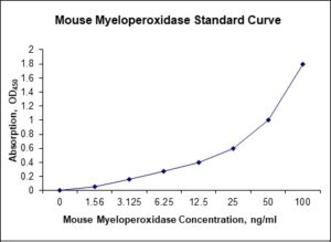
| Detection Range | 1.56-100 ng/mL |
| Specificity | Mouse MPO |
| Cross-Reactivity | < 0.5% cross-reactivity observed with available related molecules, < 50% cross-species reactivity observed with species tested. |
| Interference | No significant interference observed with available related molecules |
| Storage/Stability | 4 ºC for up to 6 months |
| Usage | For Laboratory Research Use Only. Not for diagnostic or therapeutic use. |
| Additional Notes | The kit allows for use in multiple experiments. |
Standard Curve
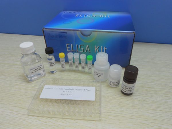
Kit Components
1. Pre-coated 96-well Microplate
2. Biotinylated Detection Antibody
3. Streptavidin-HRP Conjugate
4. Lyophilized Standards
5. TMB One-Step Substrate
6. Stop Solution
7. 20 x PBS
8. Assay Buffer
Other Materials Required but not Provided:
1. Microplate Reader capable of measuring absorption at 450 nm
2. Log-log graph paper or computer and software for ELISA data analysis
3. Precision pipettes (1-1000 µl)
4. Multi-channel pipettes (300 µl)
5. Distilled or deionized water
Protocol Outline
1. Prepare all reagents, samples and standards as instructed in the datasheet.
2. Add 100 µl of Standard or samples to each well and incubate 1 h at RT.
3. Add 100 µl of Working Detection Antibody to each well and incubate 1 h at RT.
4. Add 100 µl of Working Streptavidin-HRP to each well and incubate 20 min at RT.
5. Add 100 µl of Substrate to each well and incubate 5-30 min at RT.
6. Add 50 µl of Stop Solution to each well and read at 450 nm immediately.
Background:
Myeloperoxidase (MPO) is a member of the XPO subfamily of peroxidase that in Monkeys is encoded by the MPO gene on chromosome 17. MPO is most abundantly expressed in neutrophil granulocytes and is a lysosomal protein stored in azurophilic granules of the neutrophil and released into the extracellular space during degranulation.[1] It produces hypohalous acids to carry out their antimicrobial activity. It requires heme as a cofactor. Furthermore, it oxidizes tyrosine to tyrosyl radical using hydrogen peroxide as an oxidizing agent.[2] Hypochlorous acid and tyrosyl radical are cytotoxic, so they are used by the neutrophil to kill bacteria and other pathogens.[3] However, this hypochlorous acid may also cause oxidative damage in host tissue. Moreover, MPO oxidation of apoA-I reduces HDL-mediated inhibition of apoptosis and inflammation.[4] In addition, MPO mediates protein nitrosylation and the formation of 3-chlorotyrosine and dityrosine crosslinks. Recent studies have reported an association between elevated myeloperoxidase levels and the severity of coronary artery disease.[5] And Heslop et al. reported that elevated MPO levels more than doubled might increase the risk for cardiovascular mortality over a 13-year period.[6] It has also been suggested that myeloperoxidase plays a significant role in the development of the atherosclerotic lesion and rendering plaques unstable.[7] MPO could serve as a sensitive predictor for myocardial infarction in patients presenting with chest pain.[8] The 2010 Heslop et al. study reported that measuring both MPO and CRP provided added benefit for risk prediction than just measuring CRP alone.[6]
References
- Kinkade JM, et al. (1983). Biochem Biophys Res Communications. 114 (1): 296–303.
- Heinecke JW, et al. (1993). The Journal of Clinical Investigation. 91 (6): 2866–72.
- Hampton MB, et al. (1998). Blood. 92 (9): 3007–17.
- Shao B, et al. (2010). Chemical Research in Toxicology. 23 (3): 447–54.
- Zhang R, et al. (2001). JAMA. 286 (17): 2136–42.
- Heslop CL, e tal. (2010). J American College of Cardiology. 55 (11): 1102–9.
- Nicholls SJ, et al. (2005). Arteriosclerosis,Thrombosis and Vascular Bio. 25: 1102–11.
- Brennan ML, et al. (2003). The New England Journal of Medicine. 349 (17): 1595–604.
Be the first to review “Nori Mouse MPO ELISA Kit”
You must be logged in to post a review.




Reviews
There are no reviews yet.