


Nori Human suPAR ELISA Kit

$508.00 – $916.00
This ELISA kit is for quantification of suPAR in human. This is a quick ELISA assay that reduces time to 50% compared to the conventional method, and the entire assay only takes 3 hours. This assay employs the quantitative sandwich enzyme immunoassay technique and uses biotin-streptavidin chemistry to improve the performance of the assays. An antibody specific for suPAR has been pre-coated onto a microplate. Standards and samples are pipetted into the wells and any suPAR present is bound by the immobilized antibody. After washing away any unbound substances, a detection antibody specific for suPAR is added to the wells. Following wash to remove any unbound antibody reagent, a detection reagent is added. After intensive wash a substrate solution is added to the wells and color develops in proportion to the amount of suPAR bound in the initial step. The color development is stopped, and the intensity of the color is measured.
Alternative names for suPAR: The Urokinase receptor, urokinase-type plasminogen activator receptor (uPAR), CD87 (Cluster of Differentiation 87),
This product is for laboratory research use only not for diagnostic and therapeutic purposes or any other purposes.
- Description
- How Elisa Works
- Product Citations
- Reviews (0)
Description
Nori Human suPAR ELISA Kit Summary
Alternative names for suPAR: The Urokinase receptor, urokinase-type plasminogen activator receptor (uPAR), CD87 (Cluster of Differentiation 87),
| Assay Type | Solid Phase Sandwich ELISA |
| Format | 96-well Microplate or 96-Well Strip Microplate |
| Method of Detection | Colorimetric |
| Number of Targets Detected | 1 |
| Target Antigen Accession Number | Q03405 |
| Assay Length | 3 hours |
| Quantitative/Semiquantitative | Quantitative |
| Sample Type | Plasma, Serum, Cell Culture, Urine, Cell/Tissue Lysates, Synovial Fluid, BAL, |
| Recommended Sample Dilution (Plasma/Serum) | No dilution for sample <ULOQ; sufficient dilution for samples >ULOQ |
| Sensitivity | 12 pg/mL |
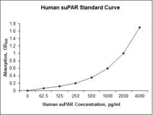
| Detection Range | 62.5-4000 pg/mL |
| Specificity | Natural and recombinant human suPAR |
| Cross-Reactivity | < 0.5% cross-reactivity observed with available related molecules, < 50% cross-species reactivity observed with species tested. |
| Interference | No significant interference observed with available related molecules |
| Storage/Stability | 4 ºC for up to 6 months |
| Usage | For Laboratory Research Use Only. Not for diagnostic or therapeutic use. |
| Additional Notes | The kit allows for use in multiple experiments. |
Standard Curve
Kit Components
1. Pre-coated 96-well Microplate
2. Biotinylated Detection Antibody
3. Streptavidin-HRP Conjugate
4. Lyophilized Standards
5. TMB One-Step Substrate
6. Stop Solution
7. 20 x PBS
8. Assay Buffer
Other Materials Required but not Provided:
1. Microplate Reader capable of measuring absorption at 450 nm
2. Log-log graph paper or computer and software for ELISA data analysis
3. Precision pipettes (1-1000 µl)
4. Multi-channel pipettes (300 µl)
5. Distilled or deionized water
Protocol Outline
1. Prepare all reagents, samples and standards as instructed in the datasheet.
2. Add 100 µl of Standard or samples to each well and incubate 1 h at RT.
3. Add 100 µl of Working Detection Antibody to each well and incubate 1 h at RT.
4. Add 100 µl of Working Streptavidin-HRP to each well and incubate 20 min at RT.
5. Add 100 µl of Substrate to each well and incubate 5-30 min at RT.
6. Add 50 µl of Stop Solution to each well and read at 450 nm immediately.
Background:
The Urokinase receptor, also known as urokinase-type plasminogen activator receptor (uPAR) or CD87 (Cluster of Differentiation 87), is a multidomain glycoprotein tethered to the cell membrane with a glycosylphosphotidylinositol (GPI) anchor. uPAR was originally identified as a saturable binding site for urokinase on the cell surface. uPAR consists of three different domains of the three-finger protein domain family, which also includes domains found in lymphocyte antigen 6 and in snake venom toxins of the three-finger toxin family, such as alpha-neurotoxins.[1] uPAR is a part of the plasminogen activation system, which in the healthy body is involved in tissue reorganization events such as mammary gland involution and wound healing. In order to reorganize tissue, the old tissue must be able to be degraded. An important mechanism in this degradation is the proteolysis cascade initiated by the plasminogen activation system. uPAR binds urokinase and thus restricts plasminogen activation to the immediate vicinity of the cell membrane. Thus uPAR seems to be an important player in the regulation of this process. However, the components of the plasminogen activation system are highly expressed in many malignant tumors, indicating that tumors are able to hijack the system, and use it in metastasis. Besides the primary ligand urokinase, uPAR interacts with several other proteins such as vitronectin, the uPAR associated protein (uPARAP) and the integrin family of membrane proteins. suPAR (NCBI Accession no. AAK31795) is the soluble form of uPAR. suPAR results from the cleavage and release of membrane-bound uPAR. suPAR concentration positively correlates to the activation level of the immune system and is present in plasma, urine, blood, serum, and cerebrospinal fluid. suPAR is a marker of disease severity and aggressiveness.[2] There are three different suPAR forms: suPARI-III, suPARII-III, and suPARI. suPARII-III is known to be a chemotactic agent for promoting the immune system.[2] suPAR is a biomarker for activation of the inflammatory and immune systems. suPAR levels are positively correlated with pro-inflammatory biomarkers, such as tumor necrosis factor-α, leukocyte counts, and C-reactive protein. SuPAR is associated with organ damage in various diseases.[3][4][5][6] Elevated levels of suPAR are associated with increased risk of systemic inflammatory response syndrome (SIRS), cancer, Focal segmental glomerulosclerosis, cardiovascular disease, type 2 diabetes, infectious diseases, HIV, and mortality. suPARnostic is a prognostic test used to detect suPAR levels in blood.
References
- Kessler, et al. (2017). Journal of Neurochemistry. 142: 7–18.
- Thunø, Maria; et al. (2009). Disease Markers. 27(3–4): 157–72.
- Enocsson, Helena; Sjöwall, Christopher (2015) Clinica Chimica Acta. 444: 234–241.
- Enocsson, Helena; et al. (2013). Translational Research. 162(5): 287–296.
- Sjöwall, C; et al. (2014). Translational Research. 165(6): 658–66.
- Hahm, E; et al. (2016). Nature Medicine. 23(1): 100–106.

Product Citations
Be the first to review “Nori Human suPAR ELISA Kit”
You must be logged in to post a review.

Reviews
There are no reviews yet.