


Nori Human PINK1 ELISA Kit

$461.00 – $832.00
This ELISA kit is for quantification of PINK1 in human. This is a quick ELISA assay that reduces time to 50% compared to the conventional method, and the entire assay only takes 3 hours. This assay employs the quantitative sandwich enzyme immunoassay technique and uses biotin-streptavidin chemistry to improve the performance of the assays. An antibody specific for PINK1 has been pre-coated onto a microplate. Standards and samples are pipetted into the wells and any PINK1 present is bound by the immobilized antibody. After washing away any unbound substances, a detection antibody specific for PINK1 is added to the wells. Following wash to remove any unbound antibody reagent, a detection reagent is added. After intensive wash a substrate solution is added to the wells and color develops in proportion to the amount of PINK1 bound in the initial step. The color development is stopped, and the intensity of the color is measured.
Alternative names for PINK1: PTEN-induced kinase 1
This product is for laboratory research use only not for diagnostic and therapeutic purposes or any other purposes.
- Description
- How Elisa Works
- Product Citations
- Reviews (0)
Description
Nori Human PINK1 ELISA Kit Summary
Alternative names for PINK1: PTEN-induced kinase 1
| Assay Type | Solid Phase Sandwich ELISA |
| Format | 96-well Microplate or 96-Well Strip Microplate |
| Method of Detection | Colorimetric |
| Number of Targets Detected | 1 |
| Target Antigen Accession Number | Q9BXM7 |
| Assay Length | 3 hours |
| Quantitative/Semiquantitative | Quantitative |
| Sample Type | Plasma, Serum, Cell Culture, Urine, Cell/Tissue Lysates, Synovial Fluid, BAL, |
| Recommended Sample Dilution (Plasma/Serum) | No dilution for sample <ULOQ; sufficient dilution for samples >ULOQ |
| Sensitivity | 25 pg/mL |
| Detection Range | 125-8000 pg/mL |
| Specificity | Human PINK1 |
| Cross-Reactivity | < 0.5% cross-reactivity observed with available related molecules, < 50% cross-species reactivity observed with species tested. |
| Interference | No significant interference observed with available related molecules |
| Storage/Stability | 4 ºC for up to 6 months |
| Usage | For Laboratory Research Use Only. Not for diagnostic or therapeutic use. |
| Additional Notes | The kit allows for use in multiple experiments. |
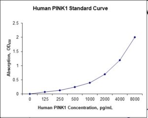
Standard Curve
Kit Components
1. Pre-coated 96-well Microplate
2. Biotinylated Detection Antibody
3. Streptavidin-HRP Conjugate
4. Lyophilized Standards
5. TMB One-Step Substrate
6. Stop Solution
7. 20 x PBS
8. Assay Buffer
Other Materials Required but not Provided:
1. Microplate Reader capable of measuring absorption at 450 nm
2. Log-log graph paper or computer and software for ELISA data analysis
3. Precision pipettes (1-1000 µl)
4. Multi-channel pipettes (300 µl)
5. Distilled or deionized water
Protocol Outline
1. Prepare all reagents, samples and standards as instructed in the datasheet.
2. Add 100 µl of Standard or samples to each well and incubate 1 h at RT.
3. Add 100 µl of Working Detection Antibody to each well and incubate 1 h at RT.
4. Add 100 µl of Working Streptavidin-HRP to each well and incubate 20 min at RT.
5. Add 100 µl of Substrate to each well and incubate 5-30 min at RT.
6. Add 50 µl of Stop Solution to each well and read at 450 nm immediately.
Background:
PTEN-induced kinase 1 (PINK1) is a mitochondrial serine/threonine-protein kinase encoded by the PINK1 gene.[1] PINK1 is synthesized as a 63 kDa protein which is often cleaved by PARL, between the 103-Alanine and the 104-Phenylalanine residues, into a 53 kDa fragment.[2] PINK1 contains an N-terminal mitochondrial localization sequence, a putative transmembrane sequence, a Ser/Thr kinase domain, and a C-terminal regulatory sequence. The protein has been found to localize to the outer membrane of mitochondria, but can also be found throughout the cytosol. PARKL1 protects cells from stress-induced mitochondrial dysfunction. PINK1 activity causes the parkin protein to bind to depolarized mitochondria to induce autophagy of those mitochondria.[3] PINK1 is processed by healthy mitochondria and released to trigger neuron differentiation.[4] Mutations in this gene cause one form of autosomal recessive early-onset Parkinson’s disease. PINK1 is intimately involved with mitochondrial quality control by identifying damaged mitochondria and targeting specific mitochondria for degradation. Healthy mitochondria maintain a membrane potential that can be used to import PINK1 into the inner membrane where it is cleaved by PARL and cleared from the outer membrane. Severely damaged mitochondria lack sufficient membrane potential to import PINK1, which then accumulates on the outer membrane. PINK1 then recruits parkin to target the damaged mitochondria for degradation through autophagy. PINK1 has been implicated in mitochondrial motility. The accumulation of PINK1 and recruitment of parkin targets a mitochondria for degradation, and PINK1 may serve to enhance degradation rates by arresting mitochondrial motility. Over-expression of PINK1 produced similar effects to silencing Miro, a protein closely associated with mitochondrial migration.[5]
References
- Valente EM, et al. (2004). Ann Neurol. 56 (3): 336–41.
- Deas E, et al. (2011). Hum. Mol. Genet. 20 (5): 867–869.
- Lazarou M, et al. (2013). Journal of Cell Biology. 200(2): 163–172.
- Dagda RK, et al. (2013). J Neurochem. 128 (6): 864–877.
- Liu S, et al. (2012). PLoS Genetics. 8 (3): e102537. doi:1371/journal.pgen.1002537.

Product Citations
Be the first to review “Nori Human PINK1 ELISA Kit”
You must be logged in to post a review.
Reviews
There are no reviews yet.