Nori Canine TLR7 ELISA Kit
$461.00 – $832.00
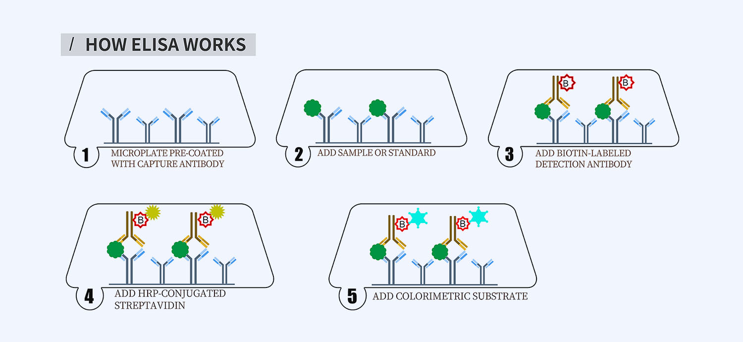
This ELISA kit is for quantification of TLR7 in dog. This is a quick ELISA assay that reduces time to 50% compared to the conventional method, and the entire assay only takes 3 hours. This assay employs the quantitative sandwich enzyme immunoassay technique and uses biotin-streptavidin chemistry to improve the performance of the assays. An antibody specific for TLR7 has been pre-coated onto a microplate. Standards and samples are pipetted into the wells and any TLR7 present is bound by the immobilized antibody. After washing away any unbound substances, a detection antibody specific for TLR7 is added to the wells. Following wash to remove any unbound antibody reagent, a detection reagent is added. After intensive wash a substrate solution is added to the wells and color develops in proportion to the amount of TLR7 bound in the initial step. The color development is stopped, and the intensity of the color is measured.
Alternative names for TLR7: Toll-like receptor 7
This product is for laboratory research use only not for diagnostic and therapeutic purposes or any other purposes.
- Description
- How Elisa Works
- Product Citations
- Reviews (0)
Description
Nori Canine TLR7 ELISA Kit Summary
Alternative names for TLR7: Toll-like receptor 7
Alternative names for canine: dog
| Assay Type | Solid Phase Sandwich ELISA |
| Format | 96-well Microplate or 96-Well Strip Microplate |
| Method of Detection | Colorimetric |
| Number of Targets Detected | 1 |
| Target Antigen Accession Number | A0A8I3SBE6 |
| Assay Length | 3 hours |
| Quantitative/Semiquantitative | Quantitative |
| Sample Type | Plasma, Serum, Cell Culture, Urine, Cell/Tissue Lysates, Synovial Fluid, BAL, |
| Recommended Sample Dilution (Plasma/Serum) | No dilution for sample <ULOQ; sufficient dilution for samples >ULOQ |
| Sensitivity | 30 pg/mL |
| Detection Range | 0.156-10 ng/mL |
| Specificity | Canine TLR7 |
| Cross-Reactivity | < 0.5% cross-reactivity observed with available related molecules, < 50% cross-species reactivity observed with species tested. |
| Interference | No significant interference observed with available related molecules |
| Storage/Stability | 4 ºC for up to 6 months |
| Usage | For Laboratory Research Use Only. Not for diagnostic or therapeutic use. |
| Additional Notes | The kit allows for use in multiple experiments. |
Standard Curve
Kit Components
1. Pre-coated 96-well Microplate
2. Biotinylated Detection Antibody
3. Streptavidin-HRP Conjugate
4. Lyophilized Standards
5. TMB One-Step Substrate
6. Stop Solution
7. 20 x PBS
8. Assay Buffer
Other Materials Required but not Provided:
1. Microplate Reader capable of measuring absorption at 450 nm
2. Log-log graph paper or computer and software for ELISA data analysis
3. Precision pipettes (1-1000 µl)
4. Multi-channel pipettes (300 µl)
5. Distilled or deionized water
Protocol Outline
1. Prepare all reagents, samples and standards as instructed in the datasheet.
2. Add 100 µl of Standard or samples to each well and incubate 1 h at RT.
3. Add 100 µl of Working Detection Antibody to each well and incubate 1 h at RT.
4. Add 100 µl of Working Streptavidin-HRP to each well and incubate 20 min at RT.
5. Add 100 µl of Substrate to each well and incubate 5-30 min at RT.
6. Add 50 µl of Stop Solution to each well and read at 450 nm immediately.
Background:
Toll-like receptor 7 (TLR7) is a protein that is encoded by the TLR7 gene.[1] TLR7 recognizes single-stranded RNA in endosomes, which is a common feature of viral genomes which are internalized by macrophages and dendritic cells. TLR7 recognizes single-stranded RNA of viruses such as HIV and HCV.[2][3] TLR7 can recognize GU-rich single-stranded RNA.[2] However, the presence of GU-rich sequences in the single-stranded RNA is not sufficient to stimulate TLR7.[3] TLR7 has been shown to play a significant role in the pathogenesis of autoimmune disorders such as lupus as well as in the regulation of antiviral immunity. The receptor TREML4 acts as an essential positive regulator of TLR7 signalling. In TREML4 -/- mice macrophages that are hyporesponsive to TLR7 agonists, macrophages fail to produce type I interferons due to impaired phosphorylation of the transcription factor STAT1 by the mitogen-activated protein kinase p38 and decreased recruitment of the adaptor MyD88 to TLR7. TREML4 deficiency reduced the production of inflammatory cytokines and autoantibodies in MRL/lpr mice, suggesting that TLR7 is a vital component of antiviral immunity and a predecessor factor in the pathogenesis of rheumatic diseases such as SLE.[4] A TLR7 agonist, Aldara, an imidazoquinoline, has been approved for topical use in treating warts caused by papillomavirus and for actinic keratoses.[5] Antiviral response elicited against avian influenza virus infection following activation of TLR7 signaling pathway is attributable to interleukin IL-1beta production.[6] Activation of TLR7 is associated with anorexia, hypoactivity, hypothermia, disturbance of feed passage in the digestive tract and the response to stress in chicks.[7] A deficient TLR7 gene caused several young patients to become seriously ill after being infected by SARS-CoV-2[8], suggesting that TLR7 plays a key role in triggering the immune response for patients of COVID-19.
References
- Yilmaz A, et al. (2005) Immunogenetics 56 (10), 743-753.
- Heil F, et al. (2004). Science. 303 (5663): 1526–9.
- Zhang Y, et al. (2016). Scientific Reports. 6: 29447.
- Ramirez-Ortiz ZG, et al. (2015). Nature Immunology. 16 (5): 495–504.
- Vender RB, Goldberg O (2005). Journal of Drugs in Dermatology. 4 (1): 58–63.
- Abdul-Cader MS, et al. (2018) BMC Res Notes 11 (1), 859.
- Tachibana T, et al. (2020) Br. Poult. Sci. 61 (3), 303-310.
- van der Made, et al. (2020). JAMA. doi:1001/jama.2020.13719
Product Citations
Be the first to review “Nori Canine TLR7 ELISA Kit”
You must be logged in to post a review.





Reviews
There are no reviews yet.