


Nori Human MIF ELISA Kit

Price range: $461.00 through $832.00
This ELISA kit is for quantification of MIF in human. This is a quick ELISA assay that reduces time to 50% compared to the conventional method, and the entire assay only takes 3 hours. This assay employs the quantitative sandwich enzyme immunoassay technique and uses biotin-streptavidin chemistry to improve the performance of the assays. An antibody specific for MIF has been pre-coated onto a microplate. Standards and samples are pipetted into the wells and any MIF present is bound by the immobilized antibody. After washing away any unbound substances, a detection antibody specific for MIF is added to the wells. Following wash to remove any unbound antibody reagent, a detection reagent is added. After intensive wash a substrate solution is added to the wells and color develops in proportion to the amount of MIF bound in the initial step. The color development is stopped, and the intensity of the color is measured.
Alternative names for MIF: Macrophage migration inhibitory factor, GLIF, MMIF
This product is for Laboratory Research Use Only not for diagnostic and therapeutic purposes or any other purposes.
- Description
- How Elisa Works
- Product Citation (0)
- Reviews (0)
Description
Nori Human MIF ELISA Kit Summary
Alternative names for MIF: Macrophage migration inhibitory factor, GLIF, MMIF
| Assay Type | Solid Phase Sandwich ELISA |
| Format | 96-well Microplate or 96-Well Strip Microplate |
| Method of Detection | Colorimetric |
| Number of Targets Detected | 1 |
| Target Antiben Accession Number | P14174 |
| Assay Length | 3 hours |
| Quantitative/Semiquantitative | Quantitative |
| Sample Type | Plasma, Serum, Cell Culture, Urine, Cell/Tissue Lysates, Synovial Fluid, BAL, |
| Recommended Sample Dilution (Plasma/Serum) | No dilution for sample <ULOQ; sufficient dilution for samples >ULOQ |
| Sensitivity | 6 pg/mL |
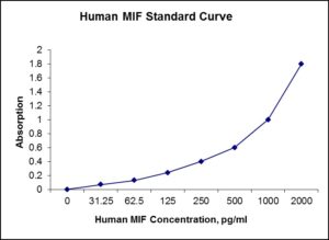
| Detection Range | 31.25-2000 pg/mL |
| Specificity | Human MIF |
| Cross-Reactivity | < 0.5% cross-reactivity observed with available related molecules, < 50% cross-species reactivity observed with species tested. |
| Interference | No significant interference observed with available related molecules |
| Storage/Stability | 4 ºC for up to 6 months |
| Usage | For Laboratory Research Use Only. Not for diagnostic or therapeutic use. |
| Additional Notes | The kit allows for use in multiple experiments. |
Standard Curve
Kit Components
1. Pre-coated 96-well Microplate
2. Biotinylated Detection Antibody
3. Streptavidin-HRP Conjugate
4. Lyophilized Standards
5. TMB One-Step Substrate
6. Stop Solution
7. 20 x PBS
8. Assay Buffer
Other Materials Required but not Provided:
1. Microplate Reader capable of measuring absorption at 450 nm
2. Log-log graph paper or computer and software for ELISA data analysis
3. Precision pipettes (1-1000 µl)
4. Multi-channel pipettes (300 µl)
5. Distilled or deionized water
Protocol Outline
1. Prepare all reagents, samples and standards as instructed in the datasheet.
2. Add 100 µl of Standard or samples to each well and incubate 1 h at RT.
3. Add 100 µl of Working Detection Antibody to each well and incubate 1 h at RT.
4. Add 100 µl of Working Streptavidin-HRP to each well and incubate 20 min at RT.
5. Add 100 µl of Substrate to each well and incubate 5-30 min at RT.
6. Add 50 µl of Stop Solution to each well and read at 450 nm immediately.
Background:
Macrophage migration inhibitory factor (MIF) was the first lymphokine/cytokine to be recognized in the pregenomics era (1). Regardless, it is one of the least understood of all inflammatory mediators (2). Human MIF is a 12.5 kDa, 115 amino acid (aa) nonglycosylated polypeptide that is synthesized without a signal sequence (3). Secretion occurs nonclassically via an ABCA1 transporter (4). The initiating Met is removed, leaving Pro as the first amino acid. The molecule consists of two α-helices and six β-strands, four of which form a β-sheet. The two remaining β-strands interact with other MIF molecules, creating a trimer (1). Structure-function studies suggest MIF is bifunctional with segregated topology. The N- and C-termini mediate enzyme activity (in theory). Phenylpyruvate tautomerase activity has been demonstrated and is dependent upon Pro at position #1 (5). Amino acids 50-65 have also been suggested to contain thiolprotein oxidoreductase activity (6). MIF has proinflammatory cytokine activity centered around aa’s 49-65. On fibroblasts, MIF induces, IL1, IL8, and MMP expression; on macrophages, MIF stimulates NO production and TNFα release following IFNγ activation (7). MIF apparently acts through CD74 and CD44, likely in some form of trimeric interaction (8). Human MIF is active on mouse cells (7). Human MIF is 90%, 94%, 95%, and 90% aa identical to mouse, bovine, porcine, and rat MIF, respectively.
References:
- Donn, R.P. and D.W. Ray (2004) J. Endocrinol. 182:1.
- Calandra, T. and T. Roger (2003) Nat. Rev. Immunol. 3:791.
- Weiser, W.Y. et al. (1989) Proc. Natl. Acad. Sci. USA 86:7522.
- Flieger, O. et al. (2003) FEBS Lett. 551:78.
- Stamps, S.L. al. (2000) Biochemistry 39:9671.
- Nguyen, M.T. et al. (2003) J. Biol. Chem. 278:33654.
- Bernhagen, J. et al. (1994) Biochemistry 33:14144.
- Leng, L. et al. (2003) J. Exp. Med. 197:1467.

Be the first to review “Nori Human MIF ELISA Kit”
You must be logged in to post a review.
Reviews
There are no reviews yet.