





















Nori Mouse TNF beta ELISA Kit
Price range: $508.00 through $916.00
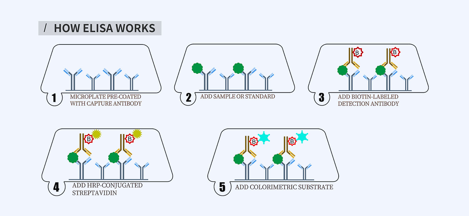
This ELISA kit is for quantification of TNF beta in mouse. This is a quick ELISA assay that reduces time to 50% compared to the conventional method, and the entire assay only takes 3 hours. This assay employs the quantitative sandwich enzyme immunoassay technique and uses biotin-streptavidin chemistry to improve the performance of the assays. An antibody specific for TNFb has been pre-coated onto a microplate. Standards and samples are pipetted into the wells and any TNFb present is bound by the immobilized antibody. After washing away any unbound substances, a detection antibody specific for TNFb is added to the wells. Following wash to remove any unbound antibody reagent, a detection reagent is added. After intensive wash a substrate solution is added to the wells and color develops in proportion to the amount of TNFb bound in the initial step. The color development is stopped, and the intensity of the color is measured.
Alternative names for TNFb: Tumor necrosis factor beta, lymphotoxin-alpha, LTA, TNFSF1
This product is for laboratory research use only not for diagnostic and therapeutic purposes or any other purposes.
- Description
- How Elisa Works
- Documents
- Product Citations (0)
- Reviews (0)
Description
Nori Mouse TNF beta ELISA Kit Summary
Alternative names for TNF beta: TNFb, tumor necrosis factor beta, lymphotoxin-alpha, LTA, TNFSF1
| Assay Type | Solid Phase Sandwich ELISA |
| Format | 96-well Microplate or 96-Well Strip Microplate |
| Method of Detection | Colorimetric |
| Number of Targets Detected | 1 |
| Target Antigen Accession Number | P09225 |
| Assay Length | 3 hours |
| Quantitative/Semiquantitative | Quantitative |
| Sample Type | Plasma, Serum, Cell Culture, Urine, Cell/Tissue Lysates, Synovial Fluid, BAL, |
| Recommended Sample Dilution (Plasma/Serum) | No dilution for sample <ULOQ; sufficient dilution for samples >ULOQ |
| Sensitivity | 6 pg/mL |
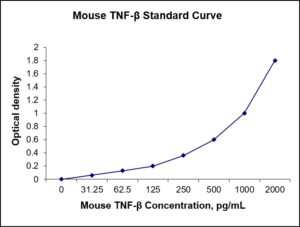
| Detection Range | 31.25-2000 pg/mL |
| Specificity | Mouse TNFb |
| Cross-Reactivity | < 0.5% cross-reactivity observed with available related molecules, < 50% cross-species reactivity observed with species tested. |
| Interference | No significant interference observed with available related molecules |
| Storage/Stability | 4 ºC for up to 6 months |
| Usage | For Laboratory Research Use Only. Not for diagnostic or therapeutic use. |
| Additional Notes | The kit allows for use in multiple experiments. |
Standard Curve
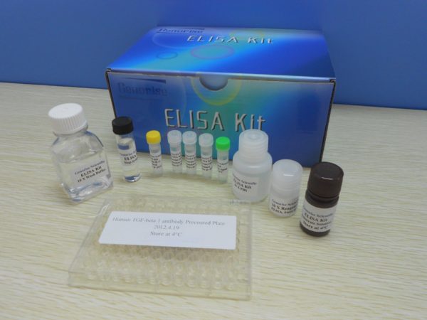
Kit Components
1. Pre-coated 96-well Microplate
2. Biotinylated Detection Antibody
3. Streptavidin-HRP Conjugate
4. Lyophilized Standards
5. TMB One-Step Substrate
6. Stop Solution
7. 20 x PBS
8. Assay Buffer
Other Materials Required but not Provided:
1. Microplate Reader capable of measuring absorption at 450 nm
2. Log-log graph paper or computer and software for ELISA data analysis
3. Precision pipettes (1-1000 µl)
4. Multi-channel pipettes (300 µl)
5. Distilled or deionized water
Protocol Outline
1. Prepare all reagents, samples and standards as instructed in the datasheet.
2. Add 100 µl of Standard or samples to each well and incubate 1 h at RT.
3. Add 100 µl of Working Detection Antibody to each well and incubate 1 h at RT.
4. Add 100 µl of Working Streptavidin-HRP to each well and incubate 20 min at RT.
5. Add 100 µl of Substrate to each well and incubate 5-30 min at RT.
6. Add 50 µl of Stop Solution to each well and read at 450 nm immediately.
Background:
Tumor necrosis factors (TNF) refer to a group of cytokines that can cause cell death (apoptosis). TNF-beta (TNF-β) also known as lymphotoxin-alpha, is a lymphokine cytokine and one of 19 identified TNF family members. TNF-β is cytotoxic for a wide range of tumor cells in vitro and in vivo.[1] TNF-β is a cytokine that is inhibited by interleukin 10. It is paracrine due to the small amounts produced. The effects are similar to TNF-alpha, but TNF-beta is also important for the development of lymphoid organs. It is a protein that is produced by Th1 type T-cells and induces vascular endothelial cells to change their surface adhesion molecules to allow phagocytic cells to bind to them. It is also known to be required for normal development of Peyer’s patches. The primary role of TNF is in the regulation of immune cells. TNF, being an endogenous pyrogen, is able to induce fever, apoptotic cell death, cachexia, inflammation and to inhibit tumorigenesis and viral replication and respond to sepsis via IL1 & IL6 producing cells. Dysregulation of TNF production has been implicated in a variety of human diseases including Alzheimer’s disease,[2] cancer,[3] major depression,[4] Psoriasis and inflammatory bowel disease (IBD). TNF can bind two receptors, TNFR1 (TNF receptor type 1) and TNFR2 (TNF receptor type 2). TNFR1 is expressed in most tissues, and can be fully activated by both the membrane-bound and soluble trimeric forms of TNF, whereas TNFR2 is found only in cells of the immune system, and respond to the membrane-bound form of the TNF homotrimer.
References
- Browning JL, et al. (1993). Cell 72 (6): 847–56.
- Swardfager W, et al. (2010). Biol Psychiatry 68 (10): 930–941.
- Locksley RM, Killeen N, Lenardo MJ (2001). Cell 104 (4): 487–501.
- Dowlati Y, et al. (2010). Biol Psychiatry 67 (5): 446–457.
Product Citations
Be the first to review “Nori Mouse TNF beta ELISA Kit”
You must be logged in to post a review.
Reviews
There are no reviews yet.