


Nori Mouse C-Met ELISA Kit

Price range: $508.00 through $916.00
This ELISA kit is for quantification of C-Met in mouse. This is a quick ELISA assay that reduces time to 50% compared to the conventional method, and the entire assay only takes 3 hours. This assay employs the quantitative sandwich enzyme immunoassay technique and uses biotin-streptavidin chemistry to improve the performance of the assays. An antibody specific for C-Met has been pre-coated onto a microplate. Standards and samples are pipetted into the wells and any C-Met present is bound by the immobilized antibody. After washing away any unbound substances, a detection antibody specific for C-Met is added to the wells. Following wash to remove any unbound antibody reagent, a detection reagent is added. After intensive wash a substrate solution is added to the wells and color develops in proportion to the amount of C-Met bound in the initial step. The color development is stopped, and the intensity of the color is measured.
Alternative names for C-Met: tyrosine-protein kinase Met, hepatocyte growth factor receptor, HGFR, proto-oncogene c-Met
This product is for laboratory research use only not for diagnostic and therapeutic purposes or any other purposes.
- Description
- How Elisa Works
- Documents
- Product Citations
- Reviews (0)
Description
Nori Mouse C-Met ELISA Kit Summary
Alternative names for C-Met: tyrosine-protein kinase Met, hepatocyte growth factor receptor, HGFR, proto-oncogene c-Met
| Assay Type | Solid Phase Sandwich ELISA |
| Format | 96-well Microplate or 96-Well Strip Microplate |
| Method of Detection | Colorimetric |
| Number of Targets Detected | 1 |
| Target Antigen Accession Number | P16056 |
| Assay Length | 3 hours |
| Quantitative/Semiquantitative | Quantitative |
| Sample Type | Plasma, Serum, Cell Culture, Urine, Cell/Tissue Lysates, Synovial Fluid, BAL, |
| Recommended Sample Dilution (Plasma/Serum) | No dilution for sample <ULOQ; sufficient dilution for samples >ULOQ |
| Sensitivity | 12 pg/mL |
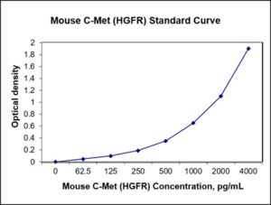
| Detection Range | 62.25-4000 pg/mL |
| Specificity | Mouse C-Met |
| Cross-Reactivity | < 0.5% cross-reactivity observed with available related molecules, < 50% cross-species reactivity observed with species tested. |
| Interference | No significant interference observed with available related molecules |
| Storage/Stability | 4 ºC for up to 6 months |
| Usage | For Laboratory Research Use Only. Not for diagnostic or therapeutic use. |
| Additional Notes | The kit allows for use in multiple experiments. |
Standard Curve
Kit Components
1. Pre-coated 96-well Microplate
2. Biotinylated Detection Antibody
3. Streptavidin-HRP Conjugate
4. Lyophilized Standards
5. TMB One-Step Substrate
6. Stop Solution
7. 20 x PBS
8. Assay Buffer
Other Materials Required but not Provided:
1. Microplate Reader capable of measuring absorption at 450 nm
2. Log-log graph paper or computer and software for ELISA data analysis
3. Precision pipettes (1-1000 µl)
4. Multi-channel pipettes (300 µl)
5. Distilled or deionized water
Protocol Outline
1. Prepare all reagents, samples and standards as instructed in the datasheet.
2. Add 100 µl of Standard or samples to each well and incubate 1 h at RT.
3. Add 100 µl of Working Detection Antibody to each well and incubate 1 h at RT.
4. Add 100 µl of Working Streptavidin-HRP to each well and incubate 20 min at RT.
5. Add 100 µl of Substrate to each well and incubate 5-30 min at RT.
6. Add 50 µl of Stop Solution to each well and read at 450 nm immediately.
Background:
c-Met, also called tyrosine-protein kinase Met or hepatocyte growth factor receptor (HGFR),[1] is a protein that is encoded by the MET gene. The protein possesses tyrosine kinase activity. MET is a receptor tyrosine kinase that is produced as a single-chain precursor. The precursor is proteolytically cleaved at a furin site to yield a highly glycosylated extracellular α-subunit and a transmembrane β-subunit, which are linked together by a disulfide bridge.[2] MET is essential for embryonic development, organogenesis and wound healing. HGF/Scatter Factor (SF) and its splicing isoform (NK1, NK2) are the only known ligands of the MET receptor. MET is normally expressed by cells of epithelial origin, while expression of HGF/SF is restricted to cells of mesenchymal origin. When HGF/SF binds its cognate receptor MET it induces its dimerization through a not yet completely understood mechanism leading to its activation. MET activation by its ligand HGF induces MET kinase catalytic activity, which triggers transphosphorylation of the tyrosines Tyr 1234 and Tyr 1235. These two tyrosines engage various signal transducers,[3] thus initiating a whole spectrum of biological activities driven by MET, collectively known as the invasive growth program. The transducers interact with the intracellular multi-substrate docking site of MET either directly, such as GRB2, SHC,[4] SRC, and the p85 regulatory subunit of phosphatidylinositol-3 kinase,[5] or indirectly through the scaffolding protein Gab1[6] Abnormal MET activation in cancer correlates with poor prognosis, where aberrantly active MET triggers tumor growth, formation of new blood vessels that supply the tumor with nutrients, and cancer spread to other organs. MET is deregulated in many types of human malignancies, including cancers of kidney, liver, stomach, breast, and brain. Normally, only stem cells and progenitor cells express MET, which allows these cells to grow invasively in order to generate new tissues in an embryo or regenerate damaged tissues in an adult. However, cancer stem cells are thought to hijack the ability of normal stem cells to express MET, and thus become the cause of cancer persistence and spread to other sites in the body. Both the overexpression of Met/HGFR, as well as its autocrine activation by co-expression of its hepatocyte growth factor ligand, have been implicated in oncogenesis.[3]
References
- Bottaro DP, et al. (1991). Science. 251 (4995): 802–4.
- Birchmeier C, et al. ().Nat. Rev. Mol. Cell Biol. 4 (12): 915–25.
- Johnson M, et al. (1995). Biochemistry and Molecular Biology International. 36 (3): 465–74.
- Pelicci G, et al. (1995). Oncogene. 10 (8): 1631–8.
- Weidner KM, et al. (1996). Nature. 384 (6605): 173–6.
- Weidner KM, et al. (1996). Nature. 384 (6605): 173–6.

Product Citations
Be the first to review “Nori Mouse C-Met ELISA Kit”
You must be logged in to post a review.
Reviews
There are no reviews yet.