


Nori Rabbit E-Cadherin ELISA Kit

Price range: $508.00 through $916.00
This ELISA kit is for quantification of E-Cadherin in rabbit. This is a quick ELISA assay that reduces time to 50% compared to the conventional method, and the entire assay only takes 3 hours. This assay employs the quantitative sandwich enzyme immunoassay technique and uses biotin-streptavidin chemistry to improve the performance of the assays. An antibody specific for E-Cadherin has been pre-coated onto a microplate. Standards and samples are pipetted into the wells and any E-Cadherin present is bound by the immobilized antibody. After washing away any unbound substances, a detection antibody specific for E-Cadherin is added to the wells. Following wash to remove any unbound antibody reagent, a detection reagent is added. After intensive wash a substrate solution is added to the wells and color develops in proportion to the amount of E-Cadherin bound in the initial step. The color development is stopped, and the intensity of the color is measured.
Alternative names for E-Cadherin: CDH1, Epithelial cadherin, Cadherin-1, CAM 120/80, uvomorulin, CD324
This product is for laboratory research use only not for diagnostic and therapeutic purposes or any other purposes.
- Description
- How Elisa Works
- Product Citation
- Reviews (0)
Description
Nori Rabbit E-Cadherin ELISA Kit Summary
Alternative names for E-Cadherin: CDH1, Epithelial cadherin, Cadherin-1, CAM 120/80, uvomorulin, CD324
| Assay Type | Solid Phase Sandwich ELISA |
| Format | 96-well Microplate or 96-Well Strip Microplate |
| Method of Detection | Colorimetric |
| Number of Targets Detected | 1 |
| Target Antigen Accession Numbe | G1TYT3 |
| Assay Length | 3 hours |
| Quantitative/Semiquantitative | Quantitative |
| Sample Type | Plasma, Serum, Cell Culture, Urine, Cell/Tissue Lysates, Synovial Fluid, BAL, |
| Recommended Sample Dilution (Plasma/Serum) | No dilution for sample <ULOQ; sufficient dilution for samples >ULOQ |
| Sensitivity | 60 pg/mL |
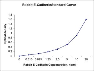
| Detection Range | 0.313-20 ng/mL |
| Specificity | Rabbit E-Cadherin |
| Cross-Reactivity | < 0.5% cross-reactivity observed with available related molecules, < 50% cross-species reactivity observed with species tested. |
| Interference | No significant interference observed with available related molecules |
| Storage/Stability | 4 ºC for up to 6 months |
| Usage | For Laboratory Research Use Only. Not for diagnostic or therapeutic use. |
| Additional Notes | The kit allows for use in multiple experiments. |
Standard Curve
Kit Components
1. Pre-coated 96-well Microplate
2. Biotinylated Detection Antibody
3. Streptavidin-HRP Conjugate
4. Lyophilized Standards
5. TMB One-Step Substrate
6. Stop Solution
7. 20 x PBS
8. Assay Buffer
Other Materials Required but not Provided:
1. Microplate Reader capable of measuring absorption at 450 nm
2. Log-log graph paper or computer and software for ELISA data analysis
3. Precision pipettes (1-1000 µl)
4. Multi-channel pipettes (300 µl)
5. Distilled or deionized water
Protocol Outline
1. Prepare all reagents, samples and standards as instructed in the datasheet.
2. Add 100 µl of Standard or samples to each well and incubate 1 h at RT.
3. Add 100 µl of Working Detection Antibody to each well and incubate 1 h at RT.
4. Add 100 µl of Working Streptavidin-HRP to each well and incubate 20 min at RT.
5. Add 100 µl of Substrate to each well and incubate 5-30 min at RT.
6. Add 50 µl of Stop Solution to each well and read at 450 nm immediately.
Background:
Epithelial cadherin (E-cadherin) also known as Cadherin-1, CAM 120/80 or uvomorulin, CD324, is a protein that is encoded by the CDH1 gene that is a tumor suppressor gene.[1] E-Cadherin is a classical member of the cadherin superfamily and is a calcium-dependent cell-cell adhesion glycoprotein composed of five extracellular cadherin repeats, a transmembrane region, and a highly conserved cytoplasmic tail. Mutations in this gene are correlated with gastric, breast, colorectal, thyroid, and ovarian cancers. Loss of E-cadherin function or expression has been implicated in cancer progression and metastasis.[2] E-cadherin downregulation decreases the strength of cellular adhesion within a tissue, resulting in an increase in cellular motility. This in turn may allow cancer cells to cross the basement membrane and invade surrounding tissues.[2] E-cadherin is also used by pathologists to diagnose different kinds of breast cancer. When compared with invasive ductal carcinoma, E-cadherin expression is markedly reduced or absent in the great majority of invasive lobular carcinomas when studied by immunohistochemistry. E-cadherin acts as an invasion suppressor and a classical tumor suppressor gene in pre-invasive lobular breast carcinoma.[3] E-cadherin transcriptional inactivation is an epi-phenomenon and part of an entire program, with much more severe effects than loss of E-cadherin expression alone”.[4] The ectodomain of E-Cadherin mediates bacterial adhesion to mammalian cells, and the cytoplasmic domain is required for internalization. E-cadherin is first expressed in the 2-cell stage of mammalian development, and becomes phosphorylated by the 8-cell stage, where it causes compaction. In adult tissues, E-cadherin is expressed in epithelial tissues, where it is constantly regenerated with a 5-hour half-life on the cell surface. Cell-cell interactions mediated by E-cadherin are crucial to blastula formation in many animals.
References
- Wong AS, Gumbiner BM (2003). The Journal of Cell Biology. 161 (6): 1191–203.
- Beavon IR (2000). European Journal of Cancer. 36 (13 Spec No): 1607–20.
- Polyak K, Weinberg RA (2009). Nature Reviews. Cancer. 9 (4): 265–73.
- Lombaerts M, et al. (2006). British Journal of Cancer. 94 (5): 661–71.

Be the first to review “Nori Rabbit E-Cadherin ELISA Kit”
You must be logged in to post a review.
Reviews
There are no reviews yet.